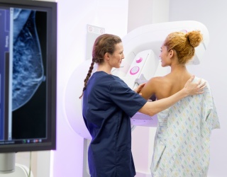
At RSNA 2025, Siemens Healthineers introduced new capabilities for its MAMMOMAT B.brilliant mammography systems, extending both contrast-enhanced mammography (CEM) and biopsy functionality. The update includes a newly developed image-reconstruction technique for contrast-enhanced examinations, designed to deliver consistent image quality and support faster diagnostic workflows within mammography.
MAMMOMAT B.brilliant already enables the generation of high-resolution 3D breast images using wide-angle tomosynthesis in five seconds. With the addition of the new ClearCEM image-reconstruction technique, the system can now support contrast-enhanced examinations without requiring a switch to other imaging modalities. The on-site solution is designed for high availability and cost efficiency, supporting long-term operational sustainability in radiology departments.
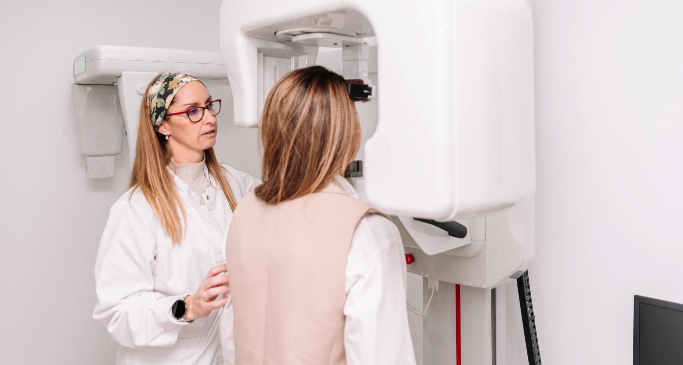
Supporting Diagnostics in Dense Breast Tissue
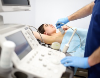
Contrast-enhanced mammography is a clinically established imaging technique used to clarify inconclusive findings or assess disease extent before surgery. By leveraging differences in iodine contrast uptake between healthy and malignant tissue, CEM supports radiologists in identifying and characterizing suspicious lesions, particularly in patients with dense breast tissue.
The new ClearCEM image-reconstruction technique is designed to provide a uniform background and consistent enhancement, supporting lesion detection and clinical decision-making. According to Siemens Healthineers, the approach may reduce the need for additional imaging and help accelerate time-to-diagnosis.
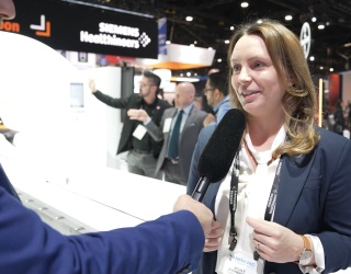
“Our goal was to empower clinicians with a solution that strengthens diagnostic confidence and broadens access to advanced imaging within the mammography workflow,” said Joanne Barry, head of X-ray Products at Siemens Healthineers Great Britain and Ireland. She emphasized that mammography remains the most accessible breast imaging modality and that expanding its capabilities can help improve access to advanced diagnostics. “Given the anxiety associated with the clarification of potential findings detected in breast cancer screening, accelerating time-to-diagnosis is essential. It reflects our commitment to innovation in women’s healthcare and to delivering personalised care.”
Integrated Contrast-Enhanced Biopsy Workflow
When a biopsy is required following a contrast-enhanced finding, the procedure is typically performed using contrast as well. ClearCEM supports this workflow by providing a contrast localizer image for tomosynthesis-guided biopsy. High depth resolution enables targeting accuracy within ±1 mm, helping maintain diagnostic precision without switching modalities.
The combination of ClearCEM-powered scout imaging and tomosynthesis-based targeting within the same compression is designed to streamline the biopsy process and increase system availability. This can be particularly relevant in high-volume clinical environments, where efficiency and throughput are critical.
Dr. Dianne Georgian-Smith, MD, Envision Healthcare, Nashville, Tennessee, USA, commented on the technology during clinical testing: “The image quality with ClearCEM is exceptional – even in dense breast tissue. ClearCEM provides a remarkably uniform background, which significantly improves the visibility of enhancing lesions.”
Focus on Workflow Efficiency and Sustainability
By integrating contrast-enhanced imaging and biopsy capabilities into a single mammography platform, Siemens Healthineers aims to support diagnostic confidence while maintaining efficient workflows. The expanded capabilities of MAMMOMAT B.brilliant are designed to help radiology practices balance diagnostic performance, system availability, and long-term economic sustainability.
Source: Siemens Healthineers