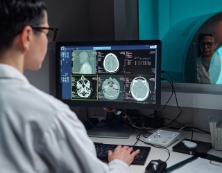
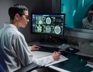
A digital twin in radiology is a virtual replica of a patient’s anatomy or imaging system that continuously integrates imaging data with other patient information. This interview takes a closer look at how digital twin technology is moving from early research in CT and MRI into clinical use across radiology and radiation oncology.
Xiaofeng Yang, PhD, DABR, professor and vice chair for medical physics research, and Chih-Wei Chang, PhD, DABR, assistant professor and medical physicist, both from the Department of Radiation Oncology at Emory University School of Medicine, share their insights on how digital twin technology is transforming imaging precision, adaptive therapy, and the future of personalized care.

One of the promises of digital twins is more personalized imaging. What are the main ways a digital twin could improve the scanning process for patients?
Digital twins can potentially make a paradigm shift in medical imaging. Specifically, a digital twin creates a virtual replica of the patient—a living, learning model that evolves with every new scan or clinical input. By combining this model with advanced physics and machine learning, we can begin to personalize the imaging process in ways that were never possible before. One major benefit is pre-scan optimization. Before a patient even enters the scanner, their digital twin can simulate how different imaging settings, such as angle, dose, or contrast level, would perform for their unique anatomy. This allows us to choose the most effective protocol upfront, reducing unnecessary exposure and avoiding repeat scans. During the scan itself, digital twins can also enable real-time adaptation. Because the virtual model continuously updates with live imaging data, the system can automatically adjust parameters to account for motion, breathing, or positioning changes, keeping the image sharp and consistent without additional scans.
Beyond improving accuracy, digital twins can help lower radiation doses, especially in repeated imaging or radiotherapy settings. By predicting exactly how much information is needed to achieve diagnostic quality, the system can minimize exposure while maintaining precision. Importantly, digital twins aren’t only for patients; they can also model the imaging equipment itself. By monitoring scanner performance in real time, these system-level twins can detect calibration drifts or performance issues before they affect image quality. The result is a smoother workflow and greater reliability across imaging sessions.
Each scan adds to the patient’s virtual record, enabling clinicians to track subtle anatomical or physiological changes over time. This creates a powerful feedback loop in which every scan becomes smarter, faster, and more tailored to the individual. In essence, digital twins shift imaging from a “one-size-fits-all” approach to a living, adaptive process that’s uniquely attuned to each patient. It’s a step toward truly personalized imaging, one where the virtual and physical worlds work together to deliver better, safer, and more precise care.
Radiology workflows are already complex, so integration is a key concern. What kind of patient data needs to be fed into the model, and how can this be integrated into routine radiology workflows?
The key to integrating digital twins is to leverage existing data, rather than adding new layers of complexity. At its core, a medical digital twin relies on three main data sources:
- Imaging data, such as CT, MRI, PET, or cone-beam CT (CBCT), which provide the anatomical and structural basis for the virtual model.
- Treatment and physiological data, such as dose maps, organ motion, or deformable registrations, which capture how the patient’s anatomy and physiology change over time.
- Clinical context, including demographics, pathology reports, and prior imaging histories, which allow the twin to learn patterns specific to each patient.
In our study, for example, we built a digital twin framework that combines planning CT and daily CBCT images to predict patient-specific setup uncertainties during proton therapy. A machine learning model, in our case, Gaussian process regression, assimilated these images to forecast how the prostate might shift between treatments. The framework then used these predictions to generate multiple treatment plans in advance, allowing clinicians to select the optimal one in real time.
This approach not only avoided re-planning delays but also fit naturally into the existing clinical workflow, using data that’s already routinely collected. That’s the crucial point: digital twin integration should feel seamless. The goal is not to replace the current workflow but to augment it, using automation, smart data fusion, and model-driven decision support. Most modern imaging systems already support automated DICOM data exchange, structured reporting, and GPU-based image registration and segmentation. These tools can feed directly into the twin without extra manual steps.
Over time, as more imaging and outcome data accumulate, the digital twin evolves into a continuously learning system, one that not only helps optimize today’s scans but also predicts tomorrow’s. The long-term vision is for digital twins to become a background intelligence layer in radiology: quietly integrating multimodal data, supporting adaptive planning, and providing real-time insight without interrupting the clinical flow. By embedding intelligence into the systems and data we already use, digital twins can make radiology workflows smarter, not heavier, a rare example where adding technology can actually simplify complexity.
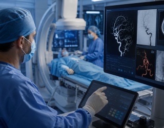
In radiation oncology, how do you see digital twins helping clinicians adjust treatment plans in real time to improve precision and patient safety?
Digital twins are redefining how we think about adaptability in radiation oncology. Traditionally, adjusting a treatment plan in real time has been limited by time, computational resources, and the need for manual intervention. Digital twins change that by acting as a continuously updated, patient-specific simulation environment, essentially a virtual copy of the patient’s anatomy and treatment system that evolves day by day.
In our research (Chang et al., 2025a; Chang et al., 2025b), we demonstrated how a digital twin could integrate planning CT and daily CBCT data to predict patient-specific setup uncertainties and generate multiple treatment plans in advance. When a patient arrives for therapy, the system evaluates the day’s CBCT in under two minutes and selects the optimal pre-approved plan, one that best fits the patient’s anatomy that day.
This avoids the need for lengthy re-optimization during treatment, which can take 30–45 minutes in traditional adaptive workflows. This is where digital twins truly enhance real-time precision and safety. Because the system already understands the patient’s anatomical variability and likely motion patterns, it can anticipate how different scenarios would affect dose distribution. The clinician can make an informed plan selection, or in future implementations, allow the system to assist in that decision, with full confidence that coverage is maintained and critical structures are protected.
From a safety standpoint, the digital twin acts as a quality assurance layer. Every daily image and dose calculation is compared against the twin’s predictive model, allowing early detection of deviations that could compromise treatment. Instead of waiting days for offline review, clinicians receive immediate feedback, reducing both underdosing to tumors and overdosing to organs at risk, such as the bladder or rectum.
In essence, digital twins can bring true adaptivity to the treatment room. They bridge the gap between planning and delivery, enabling a “plan-of-the-day” approach that’s both data-driven and patient-specific. For clinicians, this means more precise targeting; for patients, it means safer, more personalized therapy, delivered in real time, with confidence.

Ultimately, radiologists want to know how this translates into better outcomes. Have you seen any real-world cases or studies where digital twins made a noticeable difference in scan quality or efficiency?
Every innovation in radiology has to prove its value in patient outcomes, workflow efficiency, or image quality. While digital twin technology is still in its early stages, we are already beginning to see meaningful, measurable improvements in both precision and clinical efficiency.
In our recent study (Chang et al., 2025a; Chang et al., 2025b), we implemented a digital twin framework for adaptive proton therapy in prostate cancer treatment. The system used each patient’s planning CT and daily CBCT to build a personalized virtual model that predicted how the anatomy might shift from day to day. Based on those predictions, we pre-generated multiple treatment plans, each optimized for different possible anatomical scenarios.
When patients arrived for treatment, the digital twin automatically evaluated that day’s CBCT and selected the optimal plan in about two minutes, rather than the 30–45 minutes that a conventional adaptive re-optimization would require. That alone represents an improvement in efficiency and patient throughput without compromising clinical precision.
However, what’s most compelling is the impact on dosimetry and safety. Across all ten patients studied, the digital twin–based approach consistently improved or maintained tumor coverage while reducing dose to critical organs, especially the bladder neck, which is closely associated with urinary toxicity. In some cases, bladder neck doses dropped by more than 40% (Chang et al., 2025b), a clinically meaningful reduction that can directly translate to fewer side effects and better quality of life.
This is, to our knowledge, one of the first demonstrations that a digital twin framework can directly enhance both the technical quality and clinical safety of radiation therapy without adding workflow burden. While our work focused on proton therapy, the same concept can apply broadly to diagnostic imaging such as optimizing CT parameters to reduce unnecessary rescans. Even at this early stage, we are already seeing how digital twins can turn the idea of “personalized imaging” into something tangible: faster, safer, and more precise imaging decisions that directly benefit both patients and clinicians.
What are the key challenges and infrastructure requirements for implementing digital twin technology in real time, and are these achievable for most hospitals today?
The transition from research to routine clinical use is where even the best ideas are truly tested. Digital twin technology has enormous promise, but it also comes with several important challenges that we have to address thoughtfully before widespread adoption.
1. Data integration and privacy: A digital twin depends on a rich mix of clinical, imaging, and sometimes physiologic data, often coming from multiple systems and vendors. Ensuring that this data can be securely shared and synchronized, while staying compliant with HIPAA and institutional privacy rules, is essential. Most hospitals already have the building blocks: secure PACS systems, electronic health records, and DICOM-based data exchange. The key step forward is creating a safe, standardized data pipeline that feeds the digital twin automatically, without manual handling of sensitive information.
2. Computational infrastructure: Real-time or near real-time twin modeling requires substantial computing power, particularly when integrating 3D imaging and Monte Carlo–based dose calculations. In our study, for example, we used a clinical GPU server equipped with an NVIDIA Quadro RTX 8000 and dual Intel Xeon Gold CPUs hardware that’s already available in many large academic centers. With modern GPU acceleration and efficient image registration algorithms, our digital twin framework could evaluate and select the optimal treatment plan in under two minutes. So technically speaking, the infrastructure is feasible today, it just needs to be configured and scaled appropriately for clinical workloads.
3. Human factor: Radiologists, physicists, and therapists are already operating in complex, time-sensitive environments. The goal should never be to add new manual steps but to embed digital twin operations seamlessly within the existing radiotherapy or radiology workflow. That means automation, intuitive interfaces, and clear decision-support tools. Clinicians should see the digital twin as a trusted assistant, not an extra step.
There can be a cultural challenge: building trust. Clinicians need to see that digital twins are not replacing decision-making but enhancing it, offering data-driven recommendations that are transparent and clinically validated. Pilot programs, like our prostate digital twin study, are helping demonstrate that this technology can work safely within current practice standards.
Large academic centers already have the infrastructure to implement these systems. For community hospitals, cloud-based and vendor-supported models could soon make digital twin technology broadly accessible. The challenges are real, but none are insurmountable, especially given the clear clinical value. With the right infrastructure and clinical buy-in, digital twins can move from concept to clinic faster than many imagine. They represent not just a technological evolution, but a new way of thinking about continuous, patient-specific precision care.
Looking five to ten years ahead, do you see digital twins becoming standard for all advanced imaging procedures, or will adoption be limited to specific modalities and patient groups?
Looking five to ten years ahead, we think digital twins will become an integral part of precision medicine, but their adoption will evolve in phases rather than all at once. In the near term, we will likely see the fastest growth in high-value, image-guided fields, such as radiation oncology, interventional radiology, and complex MRI or CT planning, where patient-specific variability directly impacts outcomes. These are environments where the data infrastructure already exists, and where even small gains in precision can make a measurable difference in patient safety and efficiency.
As technology matures, we expect digital twins to expand into broader diagnostic imaging, integrating data from MRI, PET, ultrasound, and even wearable sensors. Imagine a future where a radiologist opens an image and the system already knows how that patient’s anatomy and physiology are expected to behave based on their digital twin. That is where we’re headed: imaging that is predictive, not just descriptive.
From a technical standpoint, the foundation is already being laid. GPU-based computing, automated image registration, and AI-driven modeling, all of which we used in our study, are becoming standard components of modern imaging systems. The next leap will come from multi-modal integration and continuous learning, where the twin evolves with each scan and clinical event, turning static imaging snapshots into a living model of patient health.
If you had to name the single biggest impact digital twins could have on radiology, what would it be?
If we had to name the single biggest impact, it would be this: digital twins will make radiology proactive rather than reactive. Instead of imaging a problem after it occurs, we will be able to anticipate how disease or anatomy is likely to change and adjust imaging or treatment strategies in advance. It’s the difference between diagnosing a patient and understanding them in motion.
Ultimately, digital twins are not just another computational tool; they represent a shift in mindset. By uniting data, physics, and machine learning into a single continuous framework, they will enable radiology to deliver on its most ambitious promise: personalized, predictive, and adaptive care for every patient.
Sources:
Chang C-W, Akkineni S S, Hu M, Shah K, Gao Y, Patel P, Jani A B, Agasthya G, Zhou J and Yang X 2025a A Digital Twin Framework for Adaptive Treatment Planning in Radiotherapy arXiv preprint arXiv:2506.14701v3
Chang C-W, Tian Z, Qiu R L J, Scott Mcginnis H, Bohannon D, Patel P, Wang Y, Yu D S, Patel S A, Zhou J and Yang X 2025b Exploration of an adaptive proton therapy strategy using CBCT with the concept of digital twins Physics in Medicine & Biology
Zhao F, Wu Y, Hu M, Chang C-W, Liu R, Qiu R and Yang X 2025 Current progress of digital twin construction using medical imaging Journal of Applied Clinical Medical Physics 26 e70226
Digital Twin Series