Scientists at the Swiss Light Source (SLS) at PSI have achieved non-destructive, high-resolution 3D mapping of brain tissue using X-rays, overcoming technical barriers that previously limited the method. The work, conducted with the Francis Crick Institute in the UK, is published in Nature Methods and sets the foundation for scaling X-ray–based neuroanatomical imaging.
Understanding Brain Connectivity Through Imaging
“The brain is one of the most complex biological systems in the world,” said Adrian Wanner, who leads the Structural Neurobiology research group at PSI. His team focuses on connectomics—mapping how neurons connect through synapses and circuits.
In just one cubic millimeter of brain tissue, there are approximately 100,000 neurons, 700 million synapses, and 4 kilometers of axonal wiring. Wanner notes the difficulty of deciphering this architecture: “If you take a neural network with 17 neurons, there are more ways to connect them than atoms in the universe, says Wanner. “So you can’t just try to model it. We need to measure it.”
Limitations of Electron Microscopy
Volume electron microscopy remains the standard for ultrastructural brain mapping, but it requires cutting tissue into tens of thousands of ultrathin slices. This process is labor-intensive, error-prone, and often results in loss of fine structure, limiting its scalability.
X-rays offer deeper penetration and the potential to image intact tissue blocks, but biological samples typically present low contrast and are prone to deformation under high-energy exposure.
A New Approach Using Radiation-Tolerant Resin and Cryogenic Imaging
To address these challenges, PSI researchers developed a method combining:
- An aerospace-grade epoxy resin that stabilizes tissue under X-ray exposure
- Cryogenic cooling to –178°C using liquid nitrogen
- A reconstruction algorithm that corrects minor deformations
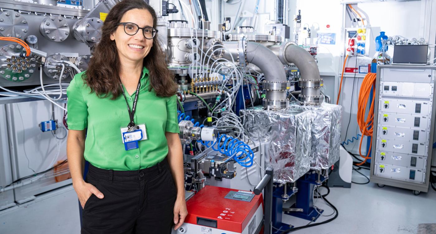
“We believe this marks a record resolution using X-ray imaging on an extended biological tissue,” said cSAXS scientist Ana Diaz. Using this method, the team imaged 10-micron-thick mouse brain samples at a 38-nanometer 3D resolution, enabling reliable identification of synapses, axons, dendrites, and other ultrastructural features.
Benefits of Coherent X-Ray Ptychography
The imaging was conducted at the cSAXS beamline using coherent X-ray ptychography. Though highly precise, current acquisition times span several days, limiting sample volume.
The recent SLS upgrade to a 4th-generation synchrotron—offering up to 100× more coherent X-ray flux—is expected to accelerate the technique.
“With one hundred times more X-ray photons hitting our sample every second, we will be able to – in principle – either image the sample one hundred times faster or image volumes one hundred times larger,” said Diaz.
Toward Larger-Scale Brain Mapping
With the technical barriers addressed and the upgraded synchrotron now operational, the researchers anticipate significantly scaling non-destructive brain tissue imaging. As Wanner summarizes: “This is not breakthrough information on the brain: it matches the best results with state-of-the-art volume electron microscopy – the current gold standard. What’s exciting is that this marks the start of what’s to come.”
Source: Nature Methods