Predicting Disease Progression Before It Happens
Osteoarthritis affects more than 500 million people globally and remains the leading cause of disability in older adults. Traditional diagnostic imaging only reveals structural damage after it has occurred, limiting physicians’ ability to predict disease progression.
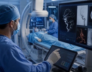
A research team at the University of Surrey (Guildford, Surrey, UK), has developed an artificial intelligence (AI) model capable of forecasting what a patient’s knee X-ray will look like one year into the future. The system provides both a visual projection and a personalized risk score, offering new insight into how osteoarthritis may evolve over time.
AI Model Trained on 50,000 Knee X-Rays
The AI was trained using nearly 50,000 knee X-rays from approximately 5,000 patients—one of the largest osteoarthritis imaging datasets to date. Using a generative diffusion model, it creates a realistic “future” version of the patient’s X-ray, highlighting 16 anatomical key points within the joint that are most likely to show degenerative change.
This feature allows clinicians to identify specific regions of concern and visualize how joint structure may deteriorate. The model operates up to nine times faster than comparable predictive tools while maintaining a compact, transparent design suitable for clinical workflows.
“Earlier AI systems could estimate the risk of osteoarthritis progression, but they were often slow, opaque and limited to numbers rather than clear images,” said Gustavo Carneiro, CVSSP Professor of AI and Machine Learning. “Our approach takes a big step forward by generating realistic future X-rays quickly and by pinpointing the areas of the joint most likely to change. That extra visibility helps clinicians identify high-risk patients sooner and personalize their care in ways that were not previously practical.”
Clinical Applications and Broader Potential
By showing current and projected images side by side, clinicians can communicate the urgency of treatment more effectively and track patient outcomes over time. The system also allows patients to visualize the potential effects of interventions such as physical therapy or lifestyle changes.
Researchers believe the same methodology could be adapted to predict disease progression in chronic conditions such as lung disease or cardiovascular disease, providing clinicians with comparable visual tools for long-term care planning.
Source: University of Surrey