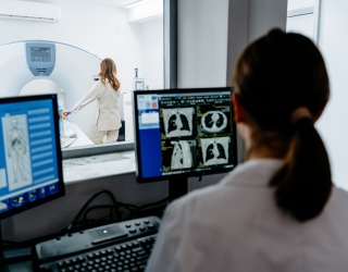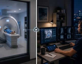
Not long ago, an MRI request for a patient with an implanted neurostimulator, pacemaker, or cochlear implant often ended in uncertainty. Device labels were misunderstood, safety information was difficult to interpret, and examinations were frequently delayed or cancelled. Today, that landscape is changing rapidly. As MR-conditional active implanted medical devices (AIMDs) become more common, medical physicists are playing an ever more central role in ensuring these examinations are both safe and clinically effective.
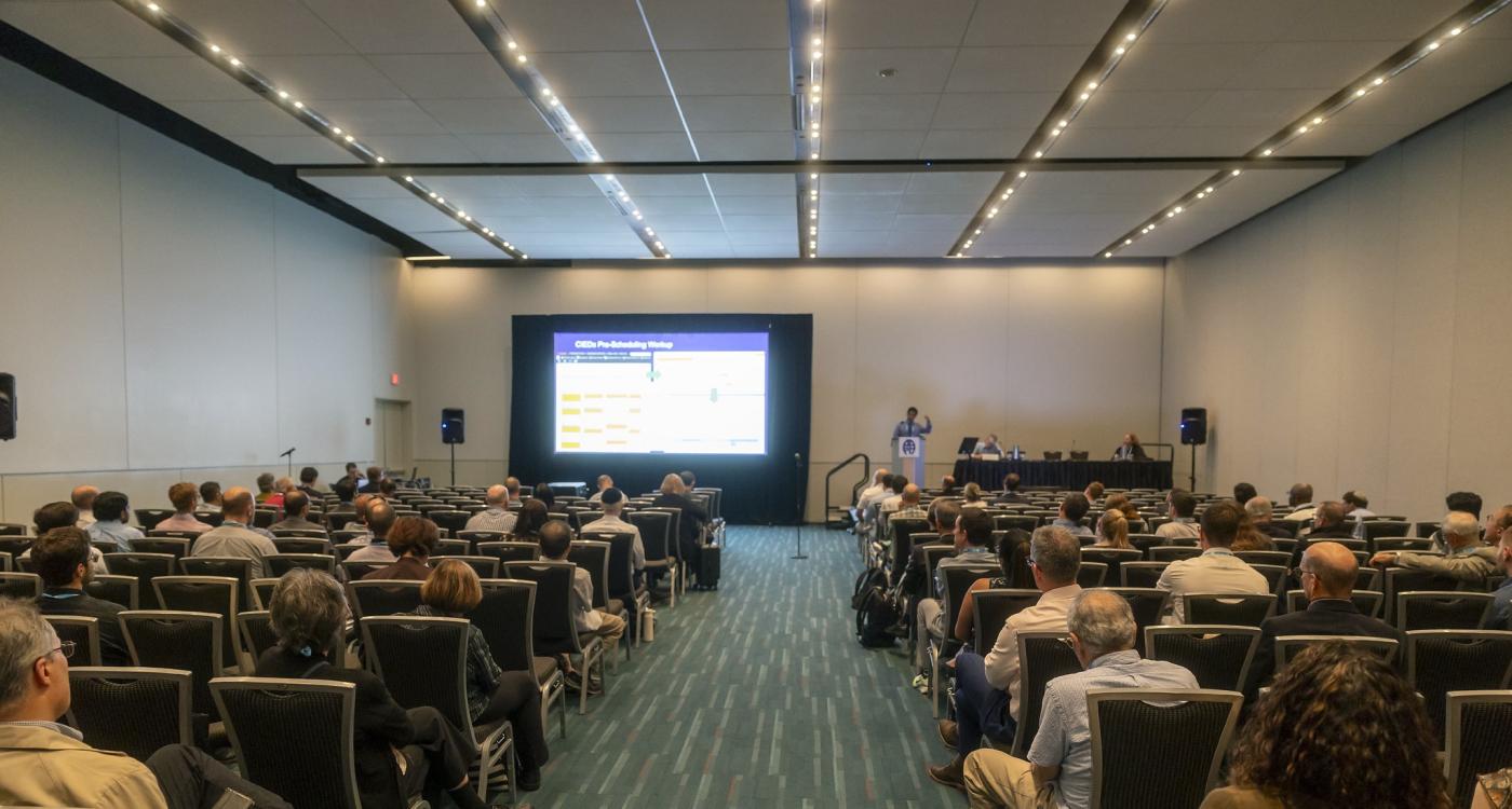
This development was at the heart of the session "MRI Safety of Active Implanted Devices: Past, Present, Future", at AAPM 2026 which explored how expanding device portfolios, new standards, and refined workflows are transforming MRI practice.
From Device Labels to Clinical Decisions
As AIMDs become routine rather than exceptional, the challenge is no longer simply identifying whether a scan is permitted but understanding under which conditions it can be performed safely and effectively. Neurostimulators, cardiac rhythm management systems, and cochlear implants each carry device-specific MRI requirements that demand interpretation beyond binary classifications. As R. Jason Stafford from the University of Texas MD Anderson Cancer Center, Houston, emphasized, “The vast majority of neurostimulants are MR conditional. Not all of them are created equal at all.”
That variability has fundamentally changed the nature of MRI safety decision-making. Instead of relying on device labels alone, clinical teams depend on specialized expertise to interpret manufacturer conditions, align them with patient-specific factors, and translate them into practical scanning protocols. Within this framework, medical physicists contribute to risk assessment, sequence optimization, and image quality preservation, ensuring that safety constraints do not unnecessarily compromise diagnostic value. In the United States, this expanding responsibility gained formal recognition with the introduction of dedicated MRI safety CPT® reimbursement codes in 2025, reflecting the growing role of medical physicists in complex implant imaging.
Scaling Expertise as Access Expands
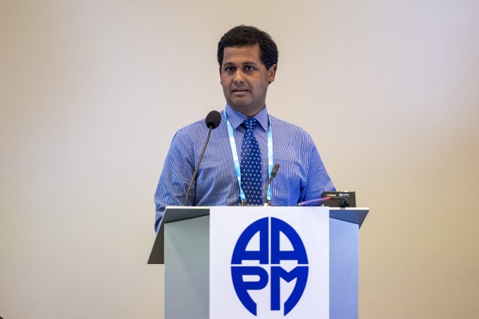
The evolution of cardiac device imaging reflects a broader shift in MRI safety. Anshuman Panda, Mayo Clinic in Phoenix, Arizona, observed, "We have shifted on the cardiac devices from, I would say, from individual risk assessment to risk assessment at scale." Rather than treating every patient with an implanted device as an exceptional case, institutions are increasingly building structured workflows that allow larger numbers of patients to be assessed and scanned safely.
What was once limited to occasional, highly scrutinized cases has evolved into a high-throughput service in many centers, supported by standardized protocols, vendor integration tools, and remote device interrogation technologies. This operational shift has been driven by close interdisciplinary collaboration, particularly between radiology and electrophysiology teams, which has enabled safe scaling without compromising oversight.
As Anshuman Panda also noted, “I don't think we would have been able to scale scanning cardiac devices if electrophysiology was not an active partner with us.”
This partnership model is increasingly being extended beyond cardiology, as other implant types present similar challenges that require coordinated input from multiple specialties. The result is a more integrated approach to patient management, where MRI safety is embedded within broader clinical pathways rather than treated as an isolated radiology concern.
Balancing Safety and Diagnostic Value
While safety remains the primary consideration, the session underscored that MRI practice cannot be reduced to risk avoidance alone. Diagnostic quality is equally critical, and implant-related factors such as artifacts, RF limitations, and positioning constraints directly influence whether an examination can answer clinical questions.
Cochlear implants illustrate this balance particularly well. Advances in magnet design, including the adoption of floating diametric magnets in newer systems, have significantly reduced both patient discomfort and procedural complexity. These developments have improved the feasibility of MRI in this patient group, yet they also highlight the ongoing challenge posed by legacy devices, which continue to require more complex preparation and careful procedural planning. As a result, informed consent and individualized risk assessment remain essential components of care.
At the same time, it was argued at the session that clinical practice is advancing faster than the evidence base. As more centers gain experience with carefully assessed off-label MRI examinations that fall outside manufacturers' labeled conditions for patients with active implants, there is a growing need to systematically document outcomes. As R. Jason Stafford noted, "We need to start publishing this experience as we use these off-label things. There's nothing in the literature to say, what's your experience with this."
From Expanding Access to Personalized MRI Pathways
Looking forward, the field is moving beyond whether patients with AIMDs can undergo MRI toward how access can be safely expanded and tailored. The increasing diversity of implantable technologies, combined with emerging evidence supporting selected off-label examinations, is driving a shift toward more nuanced, patient-specific decision-making. At the same time, developments in both low-field and ultra-high-field MRI are opening new technical possibilities that will further reshape safety frameworks.
Sustaining this progress will depend on more than technological innovation alone. Continued collaboration between clinicians, manufacturers, and medical physicists will be essential to refine guidelines, close evidence gaps, and support consistent implementation across institutions. As experience accumulates, so will the opportunity to move from broadly defined safety categories toward truly personalized MRI pathways for patients with active implants.