Neurosurgerons at UMC Utrecht, working with researchers from Eindhoven University of Technology, have introduced a method to monitor brain blood flow in real time during surgery. The technique, known as Ultrafast Power Doppler Imaging (UPDI), enables direct visualization of capillary-level perfusion, giving surgeons immediate feedback on whether brain tissue is receiving adequate blood supply.
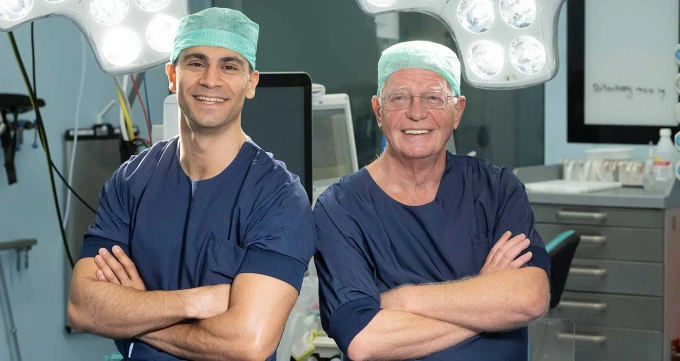
"We saw effects that would otherwise only be visible afterward, when it is unfortunately too late, for example on an MRI scan. Now, we can already measure these effects during the surgery" said Dr. Dara Niknejad, neurosurgeon at UMC Utrecht and principal investigator of the study.
Stroke Risk During Neurosurgical Procedures
Temporary blockage of blood vessels during aneurysm surgery, bypass creation, or glioma resection can lead to a high risk of stroke. This risk may start at 8% and rise to 50 % in complex cases.
Until now, surgeons had no way to observe perfusion in real time. With UPDI, they can:
- Visualize capillary-level blood flow
- Detect perfusion drops instantly
- Take immediate action to restore flow
The method was tested in a pilot study involving 10 patients, where the team continuously monitored blood flow changes during vascular procedures.
Technical Approach and Implementation
The system was developed in the Biomedical Diagnostics Lab at Eindhoven University of Technology.
"Our accurate and high-resolution measurement of blood flow in the brain allows for early detection of reduced perfusion by quantitative analysis of UPDI, such that surgeons can take timely precautions during the operation and prevent severe complications, such as cerebral infarction" said Prof. Massimo Mischi, Chair of the Signal Processing Systems Group at Eindhoven University of Technology.
Yizhou Huang, a researcher in the lab, used high-frequency ultrasound signals from multiple angles to produce detailed, high-resolution images suitable for use during neurosurgery.
Potential for Broader Clinical Use
UPDI may also be applicable in surgeries beyond neurosurgery.
"It is useful for all surgeries where continuous blood flow to the tissue is critical. We haven't tried it yet, but think about a kidney transplant, for example: the surgeon can immediately see whether the organ is fully perfused after transplantation. And even before the transplant, the organ can be assessed better with UPDI" said Niknejad.
Next Steps in Research
The research team aims to:
- Measure how accurately UPDI predicts stroke risk
- Integrate the technique more deeply into surgical workflows
- Explore use in additional surgical specialties
"Our ultimate goal is to make brain surgery safer, with fewer complications and better outcomes for patients" said Niknejad.
Source: UMC Utrecht

