Limitations of Traditional Spine Imaging
The spine’s complexity and flexibility make diagnosis challenging. While CT scans provide high-resolution images of bone and MRI captures surrounding soft tissue, both require patients to remain perfectly still. Because spinal conditions can be dynamic, static images may not reveal the full extent of problems such as deformities, fractures, or tumors.
“This work builds on a long history of using ultrasound for safe, real-time imaging, but we’re applying it in a context where it hasn’t traditionally been as effective,” said Katherine Ferrara, Professor of Radiology and Division Chief of the Molecular Imaging Program at Stanford.

Technical Advances in Ultrasound Imaging
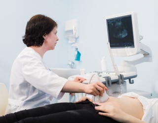
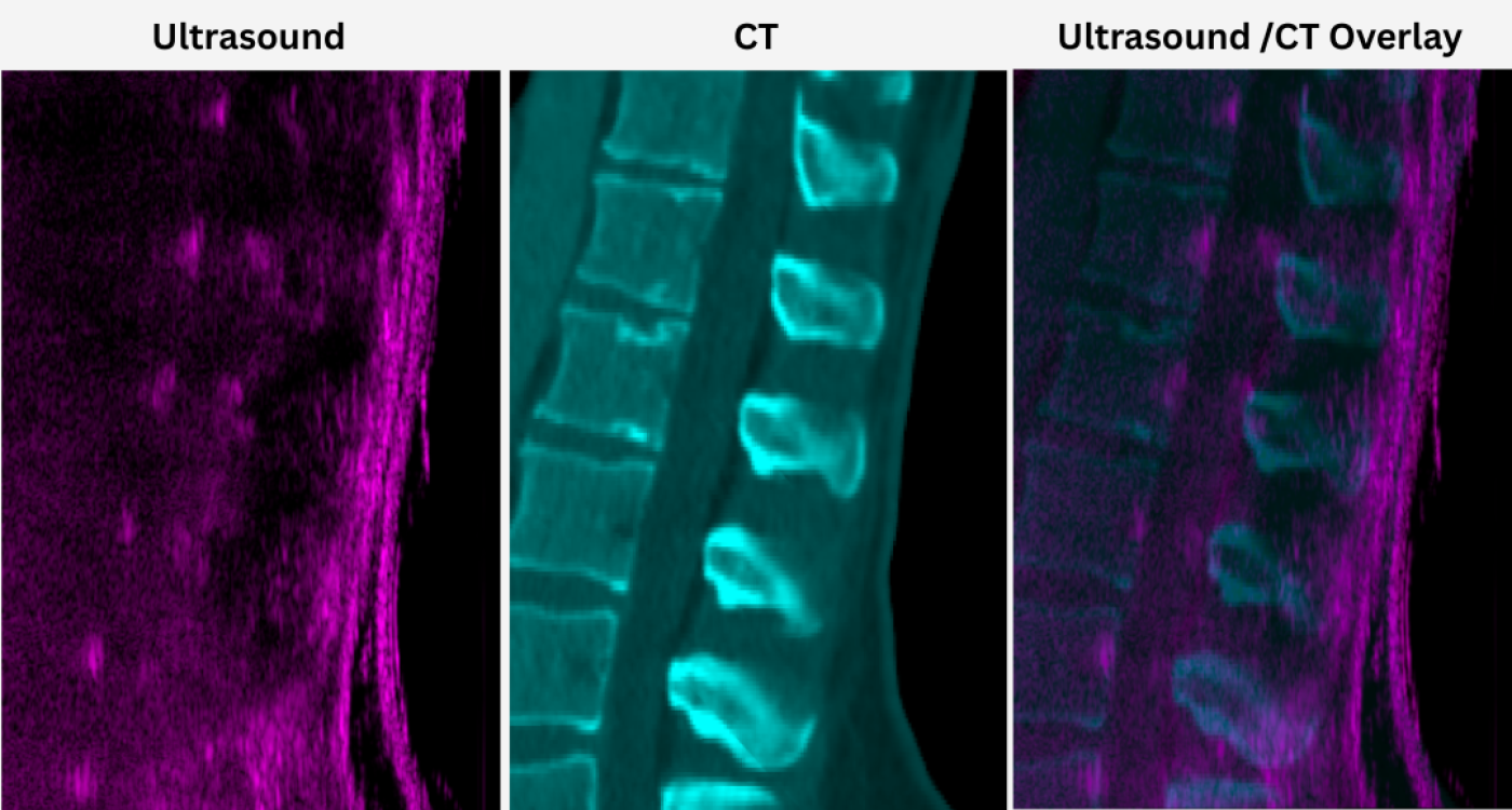
Traditional ultrasound faces limitations in spine imaging: bone blocks sound waves, and narrow probe fields restrict the view. To address this, the team developed a 9-cm-wide, 384-element transducer combined with diverging wave compounding, which transmits ultrasound waves in multiple directions.
This approach enables a panoramic, real-time view of multiple vertebrae. The study demonstrated that the device could capture images of four vertebral levels simultaneously within five seconds in healthy volunteers.
“The technical capabilities open the door to dynamic studies and procedural guidance that were simply not possible with conventional probes,” remarked Ferrara.

Clinical Applications and Procedural Guidance
For diagnostics, the large field of view allowed visualization and measurement of spinal features such as the spinous process, interspinous space, interfacet space, and spinal canal depth. Compared with CT, the ultrasound method provided similar localization without radiation exposure and at lower cost.
“Being able to visualize the spine dynamically gives us an entirely different perspective on how the spine moves and functions. For patients, this means less radiation exposure and the possibility of safer, more precise interventions, which is especially important in situations where every millimeter counts,” said Byung Chul “Jason” Yoon, Assistant Professor of Radiology at Veterans Affairs Palo Alto Health Care.
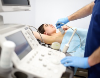
In procedural simulations, the system successfully guided a needle entering the spinal canal and tracked injected contrast in real time. The probe’s large aperture and advanced signal processing maintained image clarity across patient sizes, an important factor for guiding lumbar punctures and other interventional procedures.
Future Directions
Because it is portable and radiation-free, the system could be especially useful for bedside imaging, emergency care, or patients unable to undergo CT or MRI. It may also support dynamic studies of spinal motion and vascular flow, aiding in conditions such as scoliosis or spinal stenosis.
The research team plans further studies across diverse patient populations and clinical scenarios, as well as integration with 3D imaging capabilities.
Source: Stanford Medicine Magazine