Understanding Pulse-Synchronous Tinnitus: A Vascular Concern
Pulse-synchronous tinnitus, a form of tinnitus that pulses in time with the heartbeat, is gaining increasing attention in neuroradiology. Typically unilateral and affecting up to 5% of patients with severe tinnitus, it may be caused by vascular abnormalities in the head or neck, such as dural arteriovenous fistulas (DAVFs), venous sinus stenoses, or arteriovenous malformations (AVMs). These conditions can become dangerous if they obstruct cerebral venous outflow.
"Patients should be particularly vigilant if the ringing in the ears changes, headaches occur or even neurological deficits such as visual disturbances or dizziness are added," said Dr. Fabian Flottmann, neuroradiologist at University Medical Center Hamburg-Eppendorf (UKE)
Role of Neuroradiology in Diagnosis and Management
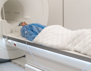
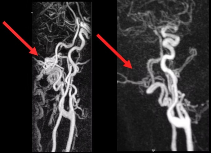
In a statement from the German Society for Neuroradiology (DGNR), neuroradiologists emphasize that their field now plays a critical role in both the diagnosis and treatment of pulse-synchronous tinnitus. Unlike conventional approaches in ENT or neurology, neuroradiological imaging allows for precise localization of vascular anomalies.
Key imaging techniques include:
- Contrast-enhanced MRI
- Time-resolved MR angiography
- CT angiography
- Catheter-based digital subtraction angiography (DSA)
These techniques help visualize abnormal flow patterns and guide therapy.
"If we identify the cause, there is a very good chance of recovery" said Dr. Fabian Flottmann
Minimally Invasive Treatment Through Image-Guided Intervention
Therapeutic options for pulse-synchronous tinnitus are typically minimally invasive and carried out via the radial (wrist) or femoral (inguinal) arteries. The specific procedure depends on the vascular condition identified:
- Fistulas are treated with embolization, using platinum coils or tissue adhesives to seal abnormal connections.
- Venous sinus constrictions may be resolved through venous stenting to improve cerebral drainage.
These image-guided interventions are designed to reduce risk and improve outcomes through targeted vascular correction.
Cross-Disciplinary Collaboration Enhances Patient Care
While pulse-synchronous tinnitus has traditionally fallen under the scope of ear, nose, and throat (ENT) medicine or neurology, neuroradiology is now central to many care pathways. The DGNR highlights the importance of interdisciplinary cooperation, especially as imaging technology and interventional capabilities advance.
At UKE Hamburg-Eppendorf, ENT specialists regularly participate in continuing education sessions offered by neuroradiologists. There is also active exchange with colleagues in private practice, reinforcing a networked approach to care.
Source: German Radiological Society