A prospective study published in Academic Radiology found higher lesion detection rates with handheld ultrasound, highlighting its continued relevance in breast imaging workflows.
Comparing Ultrasound Techniques After Breast MRI
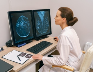
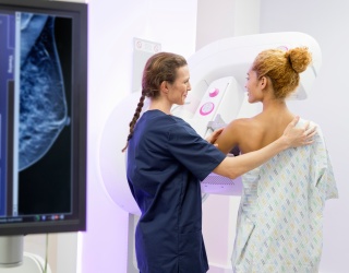
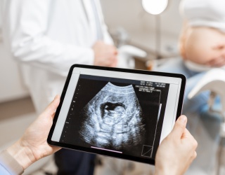
Handheld ultrasound may be more effective than automated breast ultrasound (ABUS) when performing second-look evaluations of lesions detected on breast MRI, according to new research published in Academic Radiology. The study suggests that, in this specific clinical setting, handheld ultrasound identifies a greater number of lesions, despite automation showing advantages in other imaging scenarios.
Second-look ultrasound examinations are routinely performed in women’s imaging, particularly when breast MRI reveals suspicious findings that may be more accessible to ultrasound-guided biopsy. While prior studies have reported that automated ultrasound can perform similarly to or better than handheld ultrasound in some contexts, limited research has directly compared the two techniques for second-look evaluations.
“There is limited data and no consensus on this topic in literature,” Ulku Tuba Parlakkilic, MD, with the Senology Research Institute in Istanbul, and colleagues noted.
Study Design
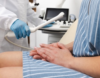
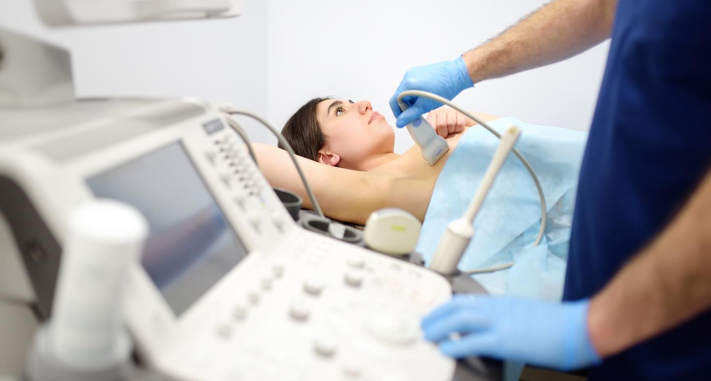
To address this gap, researchers conducted a prospective analysis involving 54 patients with a total of 66 breast lesions detected on MRI. All patients were initially referred for MRI-guided biopsy. Before undergoing biopsy, each patient received both handheld ultrasound and automated breast ultrasound examinations.
The research team compared lesion detection rates, sensitivity, and positive predictive value (PPV) for both techniques to determine whether one offered an advantage in second-look imaging.
Detection Rates and Diagnostic Performance
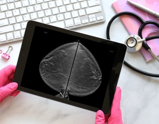
Handheld ultrasound identified 56 of the 66 lesions (84.9%), while automated ultrasound detected 46 lesions (69.7%). Of the lesions detected only by handheld ultrasound, 2 of 13 were malignant. In contrast, 2 of the 3 lesions detected exclusively by automated ultrasound were malignant.
While sensitivity was similar between the two modalities, automated ultrasound demonstrated a higher positive predictive value, exceeding handheld ultrasound by 7%. However, the authors emphasized that this advantage may not translate into clinical benefit in the second-look setting.
“We think that although a higher PPV is advantageous in the screening setting, it is not an advantage in the second look [ultrasound] setting, because not finding a lesion with [automated breast ultrasound] would lead to an MRI-guided biopsy,” the team explained. “We cannot omit a biopsy due to a negative ABUS examination, as it can have false negative results.”
The study did not identify significant differences in detection related to lesion size, depth, lesion type, location, breast density, kinetic features, or morphology.
Complementary Roles in Clinical Practice
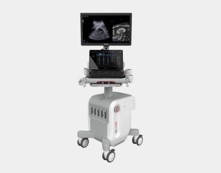
The authors acknowledged that each modality has limitations. Handheld ultrasound is highly operator dependent and typically shows lower reproducibility, while automated ultrasound reduces variability and standardizes acquisition.
“Our findings indicate that [both methods] are complementary rather than substitutive modalities, and their combined use may optimize second-look breast imaging,” the authors suggested. “Moreover, we think that [automated breast ultrasound] will and can be very helpful for radiologists with limited experience in second look US evaluation of the breast.”
Source: Academic Radiology