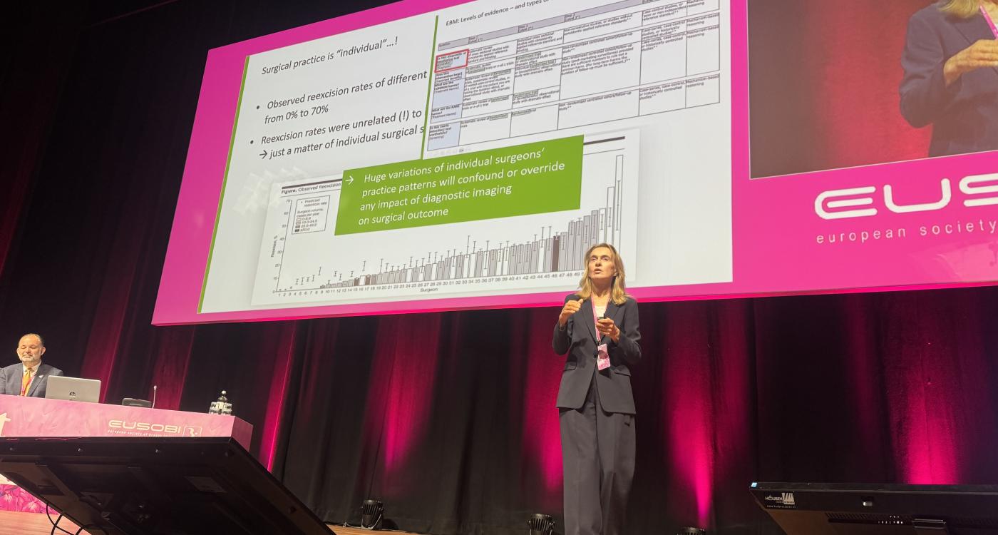
At the EUSOBI Annual Scientific Meeting 2026, Prof. Christiane Kuhl challenged one of the fundamental principles of modern clinical practice: the consistent application of evidence-based medicine.
In a keynote that combined scientific analysis with outspoken criticism, the German radiologist argued that the evidence supporting breast MRI has repeatedly been assessed differently from that supporting other imaging techniques.
“Double standards, dressed up as evidence-based medicine.”
Kuhl is director of Diagnostic and Interventional Radiology at University Hospital RWTH Aachen and president of the German Radiological Society. A pioneer of breast MRI, she has contributed extensively to research on early cancer detection, risk-adapted screening and abbreviated MRI protocols. She is also president of the European Society of Breast Imaging (EUSOBI) for the 2026–2028 term.
Her criticism therefore comes from a researcher whose work has helped establish the evidence under discussion – and from a leading representative of the European breast imaging community.
When better diagnosis is not enough
Kuhl began by distinguishing between different types of medical evidence.
Randomised controlled trials are essential when evaluating whether screening strategies improve patient outcomes, she explained. However, assessing the accuracy of a diagnostic test requires a different approach: ideally, the same patient undergoes both examinations, with results compared against a reliable reference standard.
“A randomized trial is not the highest level of evidence for every type of clinical question.”
According to Kuhl, this distinction has been insufficiently recognised in recommendations for preoperative breast MRI.
Numerous studies have demonstrated that MRI can depict the local extent of breast cancer more accurately than mammography or ultrasound, particularly when ductal carcinoma in situ (DCIS) is involved.
Nevertheless, the technique has faced demands for evidence that improved imaging reduces reoperations, recurrence or even mortality.
Kuhl questioned why such outcome requirements were imposed on breast MRI when diagnostic accuracy has been considered sufficient to justify MRI staging in other cancers, including rectal and prostate cancer.
“I'm surprised that nobody asked whether we can improve world peace by using breast MRI.”
She pointed to two earlier randomized trials, COMICE and MONET, which failed to demonstrate the anticipated surgical benefits.
Rather than accepting these findings as definitive, Kuhl questioned whether the trials adequately reflected contemporary breast MRI practice.
She argued that surgical re-excision rates vary considerably between surgeons and institutions, making them an imperfect measure of imaging performance. Furthermore, early studies lacked some of the MRI-guided localization and surgical planning techniques available today.
“You will make a good surgeon maybe somewhat better with adequate imaging information, but you can't account for a bad surgeon.”
Her underlying argument was that improved diagnostic information cannot necessarily compensate for variations in how surgeons use that information.
The debate remains clinically relevant. A subsequent systematic review and meta-analysis published in 2023 reported associations between preoperative MRI and reductions in reoperations and recurrence, although the strength of evidence varied across study designs.
The question, therefore, is not simply whether MRI identifies more extensive disease, but how reliably that information can be translated into better treatment.
Different standards for different cancers?
Kuhl drew a provocative comparison with MRI staging for rectal and prostate cancer.
In both fields, she argued, recommendations were established largely on diagnostic accuracy evidence, without requiring randomized trials demonstrating improved survival.
For breast cancer, by contrast, she suggested that the evidentiary threshold had been raised after MRI had already demonstrated superior accuracy. She also criticized the selective indications used to determine which patients should receive preoperative breast MRI.
Guidelines may recommend MRI when conventional imaging leaves uncertainty about tumor extent or when invasive lobular carcinoma is diagnosed.
But how, Kuhl asked, can clinicians use the findings of mammography to predict disease that mammography cannot detect?
“You use what mammography can see to decide whether we need to look for what it cannot see.”
Her criticism challenges not only the evidence supporting individual recommendations, but the logic of restricting a more sensitive diagnostic examination to patients whose conventional imaging already suggests a problem.
Screening: evidence accepted or ignored?
The second major part of the keynote addressed breast cancer screening. Here, Kuhl acknowledged that diagnostic accuracy alone is insufficient. A screening method must demonstrate that detecting additional cancers provides a meaningful benefit rather than merely increasing overdiagnosis.
However, she argued that breast MRI has already produced compelling evidence in several risk groups.
For women with extremely dense breasts, the Dutch DENSE trial demonstrated that supplemental MRI reduces interval cancers – cancers diagnosed between scheduled screening examinations.
Kuhl also cited evidence supporting MRI in women at intermediate familial risk, including the FaMRIsc trial, which found more favorable tumor characteristics at detection with MRI-based screening.
Yet these findings have not produced uniform recommendations across Europe. The differences are particularly evident between professional society recommendations and European Commission guidance.
EUSOBI has recommended offering MRI screening to women with extremely dense breasts. By contrast, the European Commission's ECIBC guidelines issued a conditional recommendation in July 2024 against implementing supplemental MRI in organized population-based screening for women with high breast density and negative mammography.
The Commission's recommendation reflects concerns about uncertainty in the overall balance of benefits and harms, as well as implementation considerations.
For Kuhl, however, such discrepancies illustrate a broader problem: evidence supporting breast MRI does not necessarily lead to the same clinical acceptance as evidence supporting other techniques.

A challenge to tomosynthesis
Perhaps the most contentious comparison concerned digital breast tomosynthesis (DBT).
Kuhl acknowledged that DBT detects more cancers than conventional digital mammography. But she questioned whether this improvement has been accompanied by sufficiently convincing evidence of fewer interval cancers.
She argued that additional cancer detection without a corresponding reduction in interval cancers raises concerns about overdiagnosis.
Despite this uncertainty, DBT has received favorable recommendations in European screening guidance.
Kuhl contrasted this with the more restrictive approach to supplemental breast MRI, for which randomized evidence of reduced interval cancers is available.
The comparison is provocative, although the two technologies serve different roles: DBT can replace conventional mammography within an existing screening pathway, whereas supplemental MRI introduces another examination, with additional resource requirements and potential downstream investigations.
Furthermore, the absence of a statistically significant reduction in interval cancers in individual DBT studies does not, by itself, establish that additional detected cancers represent overdiagnosis.
Nevertheless, Kuhl's criticism raises a legitimate question about how evidence, potential harms and implementation costs are weighed across different screening technologies.
Who decides what constitutes sufficient evidence?
Throughout the lecture, Kuhl returned to the distinction between generating evidence and interpreting it.
Clinical guidelines are intended to translate research into consistent patient care. But guideline committees must also make judgements about the relevance of different outcomes, the certainty of available evidence and the consequences of introducing new technologies.
Kuhl argued that these judgements have not always been applied consistently to breast MRI.
Her position is not without controversy. Greater diagnostic sensitivity does not automatically guarantee improved patient outcomes, and more extensive imaging may lead to additional biopsies, treatment changes or potentially unnecessary interventions.
Equally, evidence requirements should be proportionate to the clinical question being asked.
This tension is increasingly important as breast imaging incorporates abbreviated MRI, contrast-enhanced mammography and AI-assisted screening.
Kuhl's keynote ultimately challenged the breast imaging community to examine not only the quality of scientific evidence, but also the consistency with which that evidence influences clinical practice.
“Different clinical questions require different kinds of evidence, but the rules cannot change for different imaging methods.”
Her closing accusation – that double standards can be concealed behind the language of evidence-based medicine – was deliberately confrontational.
Whether or not one accepts her conclusions, the lecture highlighted a central issue for European breast imaging: when evidence supports a diagnostic technology, who determines whether it is sufficient – and are the same rules applied to its competitors?