AI combines micro-CT and virtual histology
Researchers at the Paul Scherrer Institute (PSI) have developed Virtual Staining of Micro-Computed Tomography (VISTACT), an AI-based platform that generates histology-like images from phase-contrast micro-computed tomography (PCµCT) data. According to the researchers, the technique combines high-resolution micro-CT imaging with machine learning to create colorized images that resemble conventional histologic stains while preserving intact tissue samples.
The method was developed to address limitations associated with conventional histology, which relies on labor-intensive preparation of thin, two-dimensional tissue sections. According to the researchers, VISTACT enables visualization of tissue architecture in three dimensions without destructive sectioning.
Machine learning translates CT data into virtual stains
The researchers trained an artificial intelligence model using paired datasets consisting of conventional histology sections and corresponding micro-CT images. A multi-stage registration workflow was used to align the histologic sections with the three-dimensional CT dataset while compensating for distortions introduced during tissue preparation.
The team then applied a conditional generative adversarial network (cGAN) to translate grayscale CT images into virtual histology images with tissue-specific color contrasts recognizable to pathologists.
According to the study, the virtual staining approach produced color patterns similar to conventional histology, including yellowish blood within small vessels, pink collagen, gray-to-violet lung surfaces, blue-violet nuclei, and dark elastic fibers.
Proof of concept in pulmonary hypertension
The proof-of-concept study focused on lung tissue samples from individuals with pulmonary hypertension. According to the researchers, CT-based virtual staining produced results comparable to laboratory histology while allowing three-dimensional mapping of remodeled pulmonary vessels.The study was published on June 17, 2026, in the Journal of The Royal Society Interface.
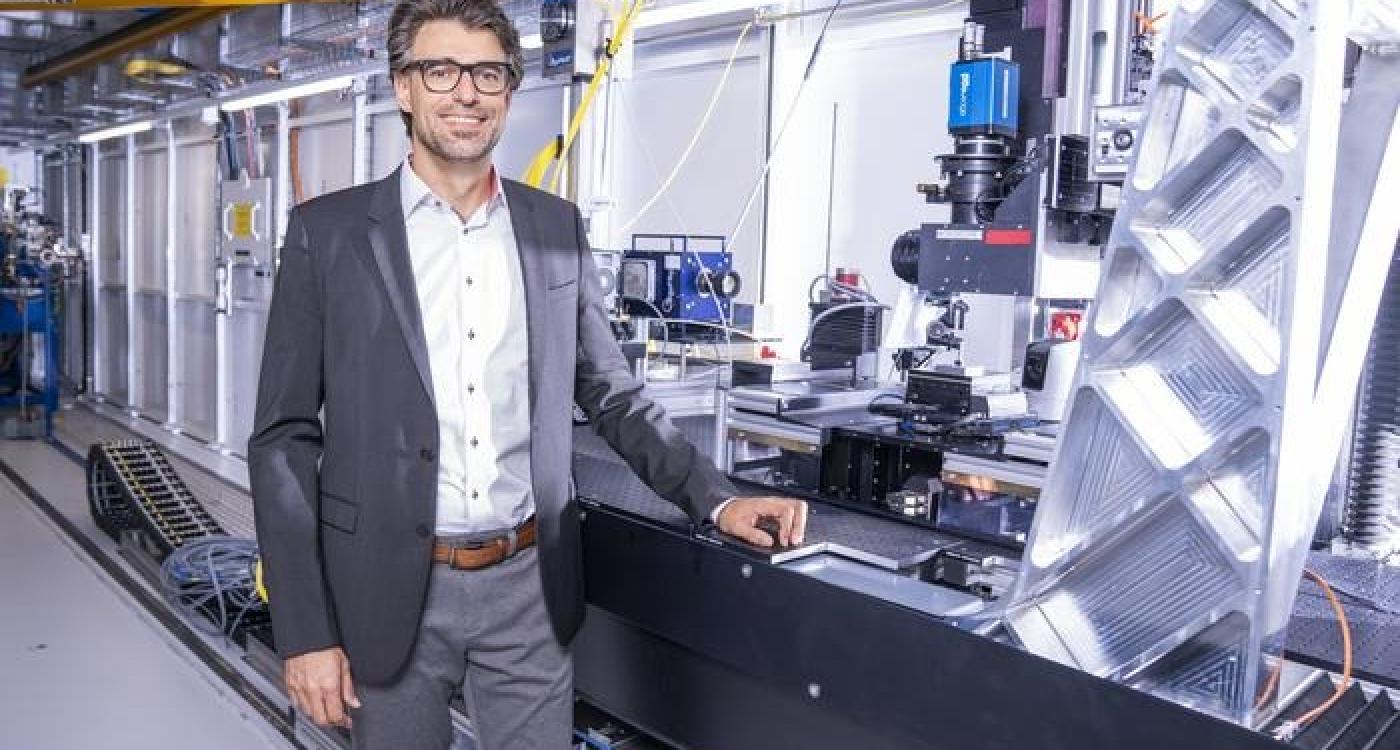
The authors also highlighted several current limitations. Phase-contrast imaging was performed at the TOMCAT beamline of the Swiss Light Source, producing very large imaging datasets. In addition, image resolution did not consistently allow visualization of individual cell nuclei. The researchers also noted that virtual staining represents statistical predictions generated by the AI model rather than direct chemical staining.
Despite these limitations, the researchers describe the method as a proof of concept for non-destructive three-dimensional pathology. They note that the approach may also be applicable to tumors, vascular lesions, and other complex tissue architectures, while potentially supporting biomarker research as imaging technology continues to develop.
Source: Paul Scherrer Institute (PSI)