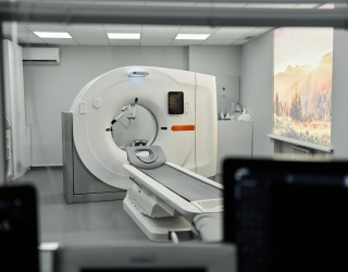
MRI is central to modern diagnosis, but its capabilities depend on more than image quality alone. Hardware design shapes what MRI can measure, while quantitative techniques can reveal biological properties beyond conventional anatomical images. Examination protocols, in turn, affect how efficiently scanners are used. Recent research across these areas explores how MRI can deliver more information while highlighting the technical and clinical challenges that remain.
10.5T MRI: Higher Field Strengths, Smarter Engineering
Ultra-high-field (UHF) MRI, generally defined as 7 T and above, provides a higher signal-to-noise ratio (SNR), which can support higher spatial resolution or shorter acquisitions. However, shorter radiofrequency (RF) wavelengths at these field strengths can cause uneven excitation, signal voids, and localized tissue heating.
A Metasurface-Based Approach
Leo Remillard and colleagues at the University of Minnesota evaluated a quadrature volume coil with a metasurface liner for 10.5T MRI, comparing it with a 16-channel parallel-transmit (pTx) array.
- Two-port design: The coil uses two ports rather than 16 independently controlled channels, potentially simplifying its construction and setup.
- Safety-related findings: Simulations showed higher safety efficiency than the reference pTx array, suggesting the potential for lower tissue heating under certain conditions.
- Remaining limitations: The prototype had lower transmit efficiency and less uniform coverage in peripheral regions than the pTx array.
The approach could offer a simpler option for selected high-field applications, although improvements in transmit efficiency and peripheral coverage are still needed. Leo Remillard from the University of Minnesota explained: "Metasurface coils are very promising for things like brain imaging at 10.5 tesla MRI. There's advantages it only has two ports versus the 16 that we compared to and that would make setup a lot easier."
Improving the hardware is one way to expand MRI's capabilities, but higher field strengths are only part of the picture. Researchers are also investigating how MRI signals can be used to characterize tissue properties that are not readily apparent in conventional anatomical images.
Mapping Tumor Physiology
Two experimental studies illustrate this shift toward quantitative MRI, examining different aspects: how tissue responds to changes in oxygen availability and whether electrical properties can provide information about cellular metabolism.
Research Spotlight 1: Oxygen-Enhanced MRI (OE-MRI)
OE-MRI measures changes in the longitudinal relaxation rate (R1) following an oxygen challenge. In a proof-of-concept study presented by Conner Ubert and colleagues at Dartmouth, a mouse model showed spatially heterogeneous tumor responses.
- Key finding: Tumor regions differed in their response to increased oxygen supply, highlighting the value of spatial imaging over single-point measurements.
- Potential relevance: Mapping these differences could support research into tumor oxygenation and hypoxia.
- Important caveat: A limited response to an oxygen challenge does not establish absolute tissue oxygen pressure or, on its own, confirm hypoxia. OE-MRI maps the tissue response to changing oxygen conditions; further validation is needed before it can be used as a routine clinical biomarker.
It highlights both the potential and the limitations of using MRI to investigate tumor biology. While OE-MRI focuses on responses to changing oxygen availability, another approach examines whether tissue electrical properties can provide complementary information about the metabolic state of tumor cells.
Research Spotlight 2: Electrical Conductivity Imaging (MREPT)
Electrical properties tomography (EPT), including magnetic resonance electrical properties tomography (MREPT), aims to estimate tissue electrical conductivity from MRI radiofrequency field information. Conductivity is influenced by factors including water content, ions and cell membranes.
Research presented by Jia Zhang investigated whether electrical conductivity is associated with tumor aggressiveness and glycolytic activity in glioma cell models.
- Key finding: In U87 glioma cells expressing the EGFRvIII mutant, direct conductivity measurements using a vector network analyzer increased from 3.86 ± 0.09 S/m to 4.90 ± 0.12 S/m compared with parental cells.
- Metabolic link: Conductivity decreased following glycolysis inhibition and increased with higher lactate concentrations.
- Potential relevance: The findings suggest that conductivity may reflect metabolic and biophysical changes associated with tumor aggressiveness. Validation in larger preclinical studies and clinical cohorts is still required.
Together, the two studies illustrate how quantitative MRI research is moving beyond structural imaging to investigate functional and biophysical characteristics of tissue. However, translating these measurements into clinical tools will require further validation.
The Overlooked Variable: Energy per Examination
Image quality and the information extracted from MRI signals are not the only considerations in developing more effective imaging. The resources required to acquire that information, particularly as healthcare providers assess operational efficiency and energy consumption.
Research by Isa Bukhari at UC Davis Health highlights substantial variation in energy use across routine MRI protocols. The study tracked energy consumption at both the examination and sequence levels at an outpatient imaging center.
Reported energy consumption per examination:
|
| ||
|---|---|---|---|
| Brain exams, 10th percentile | 7.6 kWh | ||
| Brain exams, 90th percentile | Nearly 30 kWh | ||
| Routine Brain Contrast | 22.8 kWh | ||
| MS Brain protocol | 33.8 kWh |
Note: Values refer to the protocols studied and should not be interpreted as universal MRI energy figures.
The analysis found that higher-energy examination groups also tended to have more series and longer examination times. Possible contributors include repeated acquisitions, patient motion, and protocol configuration changes. These findings identify potential targets for further investigation rather than proving that any single factor is responsible.
What Comes Next?
From high-field coil engineering to quantitative tumor imaging and energy monitoring, these studies address different stages of the MRI process. Their clinical and operational value will depend on further validation, reproducible measurements and evidence that the approaches improve clinical practice or imaging efficiency.
This article is based on the AAPM 2026 session "Emerging Frontiers in MRI Ultra-High Field Imaging, Quantitative Biomarkers, and Energy Efficiency".