Resting-State fMRI Assessed as Prognostic Tool in Traumatic Brain Injury
Recovery following traumatic brain injury (TBI) varies widely, ranging from full neurological recovery to long-term disability. Prognosis is particularly challenging in patients receiving life-sustaining therapy, where clinical signs may be limited or misleading. While resting-state functional MRI (fMRI) can capture brain activity soon after injury, its ability to predict long-term outcomes has remained uncertain.
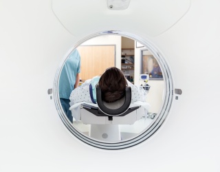
A new study led by Mass General Brigham, in collaboration with research teams across the United States and Europe, examined whether early communication between brain regions could help forecast recovery after moderate to severe TBI. The research focused on resting-state fMRI scans acquired shortly after injury and evaluated their association with functional outcomes six months later.
Analysis of Brain Network Communication
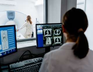
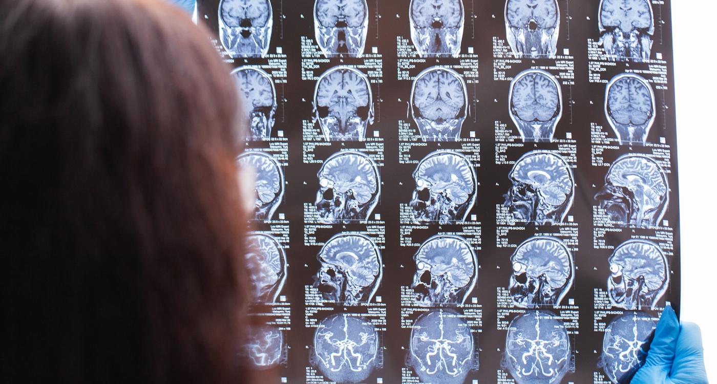
The analysis included data from three prospective patient cohorts, comprising a total of 97 individuals who underwent resting-state fMRI following TBI. Researchers evaluated functional connectivity between brain regions, including anticorrelated activity, in which activation in one area corresponds with deactivation in another. Such patterns are considered a feature of healthy brain function.
To identify predictive markers, scans from approximately half of the participants were analyzed to detect connectivity patterns linked to recovery. These findings were then tested in the remaining patients to assess their robustness. The models accounted for potential confounding factors, including sedation and level of consciousness at the time of imaging.
The researchers identified three connectivity patterns associated with favorable functional outcomes at six months. Two involved anticorrelated activity, while one showed coordinated activation across multiple regions. One of the strongest predictors involved communication between the salience network and the default mode network, which together support conscious access to information. Additional patterns linked cognitive control regions with visual processing areas, as well as connectivity between the default mode and language networks.
Consistent Performance Across Sites and Imaging Systems
The findings, published in Proceedings of the National Academy of Sciences (PNAS), demonstrated that the connectivity-based model outperformed existing prognostic tools. The results remained consistent across patients with varying injury severity, across different hospitals, and across MRI scanners used in multiple countries.
According to the study authors, the results indicate that early brain network communication plays a significant role in recovery after TBI. The use of resting-state fMRI may therefore offer a valuable tool for prognostic assessment, particularly in patients for whom clinical evaluation is challenging.
“Using brain scans, we identified signature patterns of recovery after moderate or severe TBI,” said lead author Sam Snider, MD. “These findings open new avenues for prognostic assessment in TBI, and emerging evidence suggests these patterns may be modifiable, raising the possibility of future therapeutic application.”
Future research will focus on determining whether these connectivity patterns are directly involved in healing processes and whether they can be influenced to guide treatment strategies and clinical decision-making in patients with traumatic brain injury.
Source: Mass General Brigham