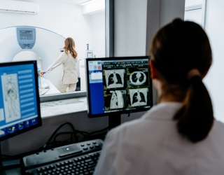
Medical imaging departments have opportunities to reduce energy consumption without necessarily disrupting clinical workflows. Much of this potential lies outside the examination itself: scanners continue to draw power between patients, during overnight periods, and while in standby or idle modes.
In a Swiss hospital study, three CT and four MR scanners consumed approximately 1.1 million kWh over one year, accounting for about 4% of the hospital's total energy consumption. Other studies have reported different proportions, with figures typically ranging from 5% to 10%, reflecting differences in measurement methods and study settings.
Studies cited in the field have also found that around 80% of scanner energy consumption can occur during idle periods. Completely powering down a scanner can reduce this demand, although this is not always practical for systems that need to remain available for emergency or short-notice examinations. Modern scanners therefore increasingly offer intermediate low-power or energy-saving modes.
Making Energy-Saving Modes Usable
The availability of energy-saving functions does not necessarily translate into their routine use. Terminology varies between vendors and even between scanner models, making it difficult for staff to identify what different modes do and when they should be used. Hannah Grover, PhD, Oregon Health & Science University, described medical physicists as sitting "at the intersection between technical scanner knowledge and clinical implementation."
Workflow considerations are another factor. Staff may be reluctant to use power-saving modes if startup times are uncertain or if switching between operating states could affect availability, calibration, or quality control. "If they don't trust it, then they're not going to use it," Grover said, referring to the importance of clinical staff confidence in the transition times.

Measurement Can Identify Where to Act
Reducing energy use also requires knowing where it is being consumed. Energy can be monitored at the scanner, examination, and sequence level, allowing departments to compare energy profiles across protocols and identify areas with high consumption or substantial variability. At UC Davis, for example, MRI protocols showed considerable variation, with a routine brain protocol having a coefficient of variation of approximately 50%. This level of measurement can also reveal opportunities for protocol optimization. In one UC Davis example, applying acceleration and deep-learning reconstruction to a T2 FLAIR protocol reduced examination time and energy consumption by approximately 60%.
MRI as a Practical Example
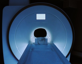
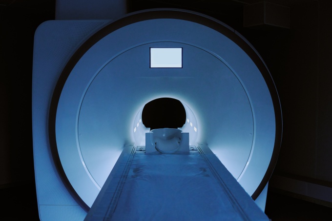
Automated shutdown and wake-up functions can reduce the need for manual intervention. In MRI, for example, modeling across four systems indicated that switching scanners off during 12-hour overnight periods could reduce overall energy. An automated power-saving mode that cycles the cryocooler can also reduce overnight consumption while keeping the system operational.
Beyond Operational Changes
Further opportunities extend to the infrastructure supporting imaging systems. Cooling can represent a substantial share of MRI energy demand; in an example, the cooling component accounted for approximately 30–40% of overall energy use. Modular chiller designs could allow cooling capacity to be scaled more closely to scanner demand. Shared electrical infrastructure across multiple scanners is another area being investigated as facilities seek to match infrastructure requirements more closely to actual combined demand.
Medical physicists can play a role in connecting these opportunities with clinical practice by measuring energy use, evaluating operating modes, and helping departments implement changes. As Grover, noted, many measures do not require major operational changes: "It just takes some small changes and education."
This article is based on the AAPM 2026 session "Optimizing Energy Use in CT and MRI: From Definitions and Measurement to Operational and Protocol Efficiency"