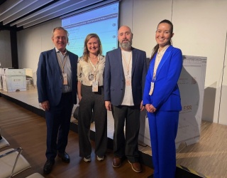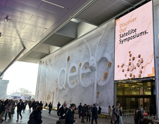
Diagnostic imaging is evolving rapidly. At the European Congress of Radiology (ECR) in Vienna, discussions focused on how diagnostic precision can be further improved while reducing the burden on patients. Presentations on new contrast agents, AI-assisted image processing, and clinical applications made it clear that progress in imaging increasingly arises from the interaction of different technologies.
One example is the AI software AiMIFY®, presented by Bracco Imaging, a global leader in diagnostic imaging. This AI-powered contrast enhancement software solution developed in collaboration with Subtle Medical, Inc. analyzes contrast-enhanced MRI scans of the brain and can selectively amplify existing contrast signals. With the commercial launch in the European Union, the technology is gaining increasing attention across imaging centers in Europe.
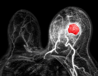
The software uses AI-based image processing to make subtle contrast enhancements more clearly visible. This allows diagnostically relevant structures to be visualized more precisely and existing image information to be used more effectively. Several presentations demonstrated that such approaches could provide additional diagnostic insights, particularly in the case of very small contrast-enhancing lesions. Post-processing signal enhancement can reveal structures that are difficult to detect in conventional reconstructions.
The goal of these approaches is not to generate new image data but to extract more diagnostic value from existing information. In complex diagnostic situations, this can help detect subtle changes earlier and classify findings more precisely.
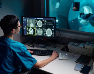
“Radiology is now a data-intensive discipline,” said Mathias Prokop, Professor of Radiology at Radboud University Nijmegen and Chair of the Department of Radiology. Modern imaging generates large volumes of complex data. The challenge, he noted, is to analyze these data in ways that produce reliable diagnostic information. Advances in reconstruction techniques and AI may help make weak signals more visible and improve the interpretation of imaging data.
Improving image quality while reducing radiation exposure
One key trend involves combining new reconstruction techniques, optimized examination protocols, and advanced software. The aim is to improve diagnostic image quality while reducing radiation exposure or contrast agent doses.

“The challenge lies in striking the right balance between image quality and patient safety,” explained Daniela Bernardi of the Department of Biomedical Sciences at Humanitas University in Milan. New technologies, she said, allow this balance to be managed far more precisely than was possible just a few years ago.
Technological advances are also transforming neuroradiological imaging. Bronwyn Hamilton, Professor of Radiology at Oregon Health & Science University, presented examples involving the detection of very small contrast-enhancing lesions. In such situations, even slight improvements in image quality can be decisive.
Hamilton presented cases in which higher contrast resolution allowed very small lesions to be delineated more clearly and diagnostic uncertainty to be reduced. “Improved image quality can directly help to detect lesions earlier and more reliably,” she said.

Ajit Shankaranarayanan, Chief Product Officer at Subtle Medical, also highlighted the potential of AI-assisted image processing. Such methods, he explained, could help extract more diagnostic information from existing image data and maintain stable diagnostic quality even when lower contrast doses are used.
These improvements may influence not only the visualization of individual structures but also the interpretation of imaging findings and clinical decision-making.
Contrast agents remain central to diagnostic imaging
Alongside advances in software and reconstruction technologies, developments in contrast agents continue to play a crucial role in modern imaging.
One example is the MRI contrast agent delivered by Bracco (gadopiclenol). Thanks to its high relaxivity, diagnostically relevant image quality can be achieved using a lower dose of gadolinium.
This physical property makes it possible to obtain strong diagnostic information while reducing exposure to the active substance. In examinations where very small structural changes must be detected, stronger contrast enhancement can be particularly important.
Josef Vymazal, Head of the Department of Radiology at Na Homolce Hospital in Prague, also highlighted this development. High-relaxivity contrast agents can help visualize very small contrast-enhancing lesions more clearly. At the same time, reduced contrast agent doses open new possibilities for improving the safety profile of diagnostic examinations.

“Innovation often arises from the combination of different technologies,” said Elena Magalotti, Global Brand Manager Digital at Bracco. Advances in contrast agents, image processing, and clinical protocols are increasingly working together to create new diagnostic opportunities.
New possibilities in pediatric imaging
Safety is another key consideration in diagnostic imaging. Regulatory developments are currently expanding the possible applications of modern contrast agents.
Andrea Rossi, Head of the Pediatric Neuroradiology Division at the Gaslini Institute in Genoa, noted that contrast agents are increasingly being approved for additional patient groups. “Extended approvals, such as for children under two years of age, open up new diagnostic possibilities,” he said.
Precise diagnostics are particularly important in pediatric imaging, while examinations must remain as gentle as possible. New contrast agents and optimized imaging protocols can help reconcile these requirements more effectively.
The interplay of multiple technologies
Discussions at the ECR showed that advances in imaging today rarely result from a single innovation. Instead, diagnostic practice evolves through the interaction of multiple technologies.
Contrast agents, AI-assisted image processing and optimized examination protocols are becoming increasingly interconnected. The goal is to extract greater value from existing diagnostic information while making imaging examinations more efficient and safer.
This development is likely to shape diagnostic imaging in the years ahead. Progress will arise less from individual technological breakthroughs than from the intelligent integration of different innovations.