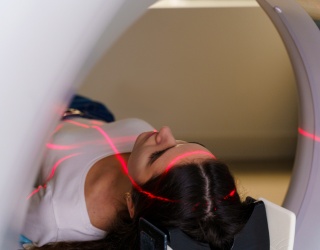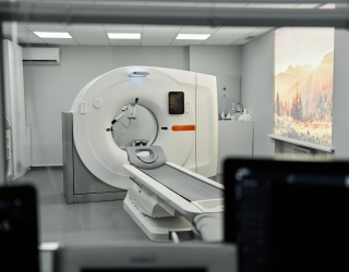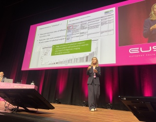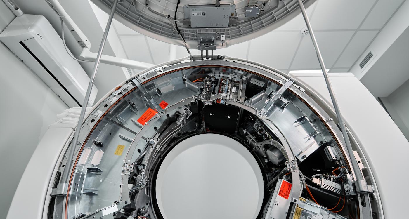
Cone-beam computed tomography (CBCT) has become an established tool in image-guided interventions and radiotherapy. Yet image artifacts, scatter, and limitations in image quality continue to restrict its capabilities. Researchers are now exploring new scanner configurations and reconstruction methods to address these challenges. Three developments illustrate how the field could move beyond conventional anatomical imaging.
New hardware for faster, more flexible imaging
Wojciech B. Zbijewski, Johns Hopkins University in Baltimore, presented two approaches to multi-source CBCT: azimuthal arrays, which position X-ray sources around the scanner, and longitudinal arrays, which arrange them along its axis of rotation. The two configurations pursue overlapping but distinct goals. Azimuthal arrays can reduce the rotation required for image acquisition and simplify the scanner's mechanical design. Longitudinal arrays, meanwhile, can help reduce cone-beam and scatter artifacts by using narrower X-ray beams.
One example was a portable brain imaging system designed for use in ambulances. It uses 21 carbon nanotube X-ray sources and a curved flat-panel detector. Rather than rotating continuously, the gantry moves between three angular positions, producing 63 projection views over approximately 200 degrees.
Zbijewski said "the objective was to make a device that is as simple mechanically as possible" to support portability. The system is designed to detect brain bleeds and, he noted, "is not only a concept": it is now undergoing early-stage clinical trials.
The limited number of projections creates a reconstruction challenge. In phantom experiments with simulated brain bleeds, diffusion posterior sampling (DPS) showed a slightly better trade-off between artifact reduction and bleed delineation than conventional model-based reconstruction.
Spectral imaging adds another dimension
Matthew W. Jacobson, Department of Radiation Oncology in Brigham and Women’s Hospital, examined how multispectral CBCT could provide information about tissue composition that conventional single-energy imaging cannot capture. By acquiring projection data at different X-ray energy spectra, spectral systems can distinguish materials and generate virtual monoenergetic images, potentially reducing beam-hardening artifacts and adjusting tissue contrast.
Jacobson highlighted applications of spectral imaging in diagnostic imaging, including distinguishing uric acid from calcium kidney stones, identifying uric acid crystal deposits associated with gout, and removing bone from CT angiography images to improve the visualization of aneurysms. These examples illustrate the broader potential of spectral imaging, although they are not all established CBCT applications.
Radiotherapy is another area of interest. Spectral imaging can support treatment planning by generating images in which iodine can either be retained or removed, depending on the clinical task. As Jacobson explained, "including iodine is beneficial for delineating tumors, whereas removing it is desirable for more accurate dose calculations."
However, bringing these capabilities into the treatment room remains challenging. "This is a much less mature area," Jacobson said, noting that treatment-room linear accelerators traditionally provide single-energy CBCT. His group is investigating multilayer detectors, software-based kV switching and combined kV/MV imaging. These approaches are at different stages of development: while some detector configurations have been tested in prototypes, the integration of multiple kV and MV spectral channels remains a long-term goal.
Addressing Reconstruction Challenges with AI
The potential of new hardware and spectral imaging also depends on reconstructing reliable images from complex measurement data. Grace Jianan Gang, University of Pennsylvania in Philadelphia, presented physics-informed diffusion posterior sampling (DPS), which combines a learned image prior with measurement data and imaging physics.
Her research explored the use of DPS for scatter correction and spectral material decomposition. In experiments using a Kyoto Kagaku phantom and a kV-switching CBCT system, jointly estimating scatter and performing material decomposition reduced cupping artifacts in water images and bias in the calcium basis images.
However, Gang cautioned that visually convincing images do not necessarily guarantee diagnostic reliability. Deep learning models can struggle to generalize across different imaging conditions and may generate artificial anatomical structures, known as hallucinations. She emphasized that improvements in reconstruction cannot replace good data acquisition: "Good data acquisition still plays an important role."
Gang also highlighted the need for more clinically meaningful evaluation methods. She advocated moving beyond conventional image-quality metrics such as PSNR and SSIM toward task-based metrics and methods to quantify hallucinations.
From prototypes to clinical evaluation
Across the three presentations, the developments ranged from systems already entering early clinical trials to experimental reconstruction methods and treatment-room prototypes. Together, they highlight different ways to address current CBCT limitations, from scanner design and material differentiation to image reconstruction. Their clinical value will ultimately depend on demonstrating reliable image quality and measurable benefits for diagnosis and treatment.
This article is based on the AAPM 2026 session "Emerging Technologies in Cone Beam CT".