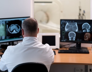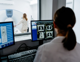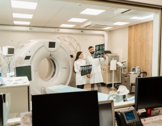
For radiographers, learning how to use an MRI or CT scanner is only part of the job. The harder part often begins when the patient enters the room. No two examinations are exactly the same. Patients breathe differently, their anatomy varies, contrast behaves differently depending on their physiology, and cardiac function can change the timing of an acquisition. That unpredictability is part of everyday clinical practice — but it is difficult to recreate in training.
This is where digital humans could add a new layer to simulation. Instead of presenting learners with a fixed virtual patient, digital humans can be programmed to behave differently. Their respiratory cycle, heart rate, stroke volume or response to contrast can change from one case to another. The idea is to make trainees react to the patient rather than simply follow a set of steps.
Matthew Hayes, President and creator of ScanLab, says this was the problem the company kept running into while trying to make MRI and CT simulation more realistic. “It’s the humans that make every exam different, every exam difficult, every exam special.” That observation shaped ScanLab’s move towards digital patients. The company has developed virtual human models with different body types and physiological characteristics and is working to make their internal behavior more closely resemble that of real patients.
Making the Patient Unpredictable
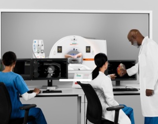
The difference becomes particularly relevant in examinations where timing matters. In a simulated CT pulmonary angiography exam, for example, the learner has to locate the pulmonary artery, place the region of interest correctly and time the scan as contrast moves through the virtual patient. Breathing can also be programmed, meaning that the patient moves during the examination rather than remaining completely static.
That creates something trainees cannot easily get in clinical practice: the freedom to get it wrong. A student can scan too early, too late or choose the wrong position and see what happens without exposing a patient to unnecessary radiation or wasting a real contrast injection. They can then repeat the case until they understand not only what to do, but why.
Learning to Adapt, Not Memorize
For Hayes, that opportunity to practice is closely linked to confidence. “Mentorship and confidence is what changes someone,” he said, describing the value of giving learners more opportunities to encounter difficult examinations before facing them in clinical practice. There is also a wider educational point. A simulator becomes less useful if learners can predict exactly what will happen every time. If the same amount of contrast always reaches the same vessel at the same second, students may simply learn the pattern.
Introducing patient variability makes that harder. Learners must understand the principles behind the examination and adapt when conditions change. That may ultimately be where digital humans have the most value. The goal is not to make simulation impressive for the sake of it. It is to recreate the parts of clinical practice that equire judgement, adaptation and experience. If simulation can do that, it moves one step closer to preparing radiographers for the part of the job that cannot be learned from a protocol alone.
This article is based on presentations and statements from the RSNA 2025 session “Digital Humans: A New Level of Clinical Realism in Simulation - presented by ScanLabMR/ScanLabCT”.