Dual Imaging Approach Addresses Diagnostic Gap in MINOCA
Patients presenting myocardial infarction but no obstructive coronary artery disease (MINOCA) often leave without a clear diagnosis. According to findings reported by MedImaging International, a combined imaging strategy using coronary optical coherence tomography (OCT) and cardiac magnetic resonance imaging (MRI) can help determine the underlying cause in most cases.
The results were presented at the American College of Cardiology’s 2026 Annual Scientific Session and published in Circulation.
OCT and Cardiac MRI Provide Complementary Insights
The dual imaging protocol integrates two complementary modalities:
- Coronary OCT: High-resolution intravascular imaging to detect plaque disruption or thrombus not visible on angiography
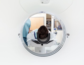
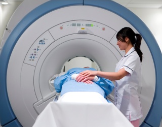
- Cardiac MRI: Assessment of myocardial injury patterns to differentiate ischemic from nonischemic causes
This combined approach allows clinicians to evaluate both coronary artery pathology and myocardial tissue characteristics.
Study Results from the HARP Trial
The Heart Attack Research Program (HARP), an international prospective study across 28 sites in the United States, Canada, and the United Kingdom, included 336 patients with MINOCA.
Key findings include:
- 79% diagnostic yield using combined OCT and MRI
- 59% ischemic causes, including plaque pathology, vasospasm, or clotting
- 20% nonischemic conditions, such as myocarditis, takotsubo syndrome, or other cardiomyopathies
The study population had a median age of 58 years and included 270 women and 66 men. Researchers reported that diagnostic yield was higher with the combined imaging protocol than with either modality alone.
Implications for Clinical Practice
The findings highlight the limitations of standard coronary angiography, which cannot detect intramural plaque abnormalities or subtle myocardial injury. Clinical characteristics, biomarkers, and initial imaging were not sufficient to predict which patients would benefit from a specific modality.
“When arteries are not badly blocked, it can be unclear what caused the event. What we show is that in most cases, we can find the underlying explanation, and most often it is a true heart attack. Our results support the need to do specialized imaging in all patients with MINOCA, because we could not reliably predict who will have specific imaging findings,” said Harmony R. Reynolds, MD, director of the Cardiovascular Clinical Research Center, Leon H. Charney Division of Cardiology, NYU Langone Health.
“We had hoped to be able to tailor testing to individual patients. Instead, we found that comprehensive imaging is often necessary to get the full answer,” added Dr. Reynolds.
Relevance for Radiology and Cardiac Imaging
The study reinforces current guideline recommendations for additional imaging in MINOCA and supports a multimodality imaging approach in cardiac diagnostics. By improving diagnostic clarity, the combined use of OCT and cardiac MRI may support more targeted treatment decisions and post-discharge management.