Phase II Study Explores Molecular Imaging for Endometriosis Detection
Researchers from the University of Oxford and Serac Healthcare Limited have reported Phase II findings suggesting that the molecular imaging agent 99mTc-maraciclatide may support non-invasive detection and monitoring of endometriosis.
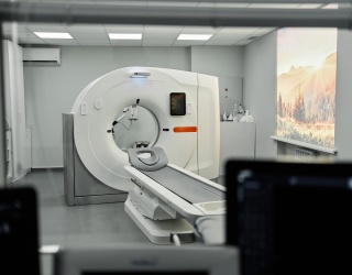
The results, published in The Lancet Obstetrics and Gynaecology and reported by MedImaging International, focused on the use of single-photon emission computed tomography-computed tomography (SPECT-CT) imaging for visualizing endometriosis lesions, including superficial peritoneal endometriosis, which is frequently difficult to identify using conventional imaging techniques.
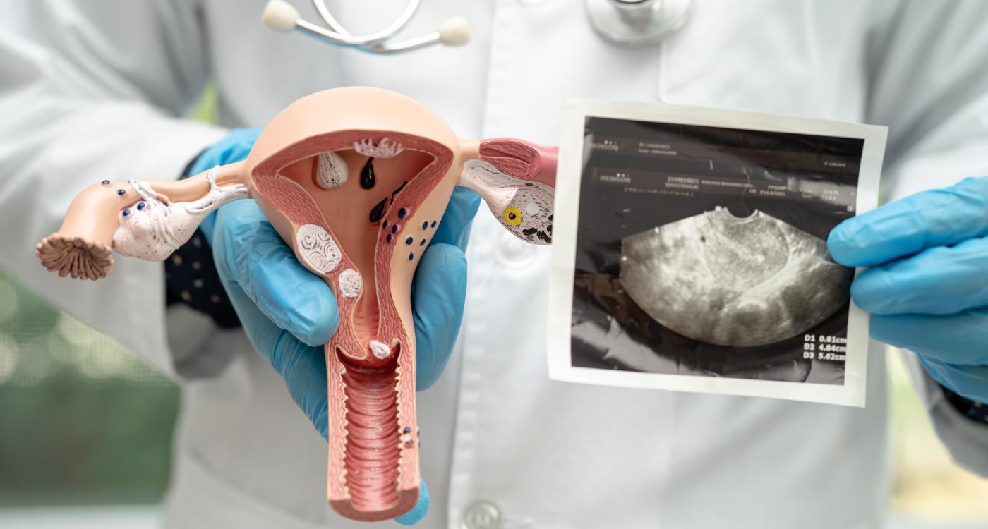
According to the researchers, current diagnosis of endometriosis often requires laparoscopic surgery because standard imaging methods may fail to detect certain lesion types. The new molecular imaging approach was developed to support lesion visualization and longitudinal disease assessment.
SPECT-CT Imaging Evaluated in DETECT Study
The exploratory Phase II DETECT study included 20 participants with suspected or confirmed pelvic or thoracic endometriosis. Patients underwent SPECT-CT imaging following intravenous administration of 99mTc-maraciclatide, a gamma-emitting radiotracer designed to bind the αvβ3 integrin associated with angiogenesis, a cardinal feature of inflammatory diseases.
Researchers compared imaging findings with surgical reports and lesion localization to evaluate concordance between imaging and operative findings.
According to the study:
- Imaging findings matched surgical results in 16 of 19 evaluable cases
- Lesions were visualized in 14 of 17 surgically positive participants
- Two cases of thoracic endometriosis were identified
- No false positives were reported
- The tracer detected lesions across all endometriosis subtypes
The study authors also noted that the imaging agent was well tolerated and demonstrated high patient acceptability.
Potential Role in Superficial Peritoneal Endometriosis
The researchers highlighted findings related to superficial peritoneal endometriosis, the most common subtype of the disease and one that is often difficult to detect with ultrasound or MRI.
Among 10 participants with superficial peritoneal endometriosis who previously underwent transvaginal ultrasound and/or MRI within 12 months, none of the conventional imaging examinations identified the disease.
“These exciting findings indicate that maraciclatide offers a highly promising diagnostic and monitoring tool, particularly for superficial peritoneal endometriosis, which is the most common and yet the hardest type of endometriosis to identify,” said Dr. Tatjana Gibbons, lead author on the paper and investigator on the study from the Nuffield Department of Women’s and Reproductive Health, University of Oxford.
The researchers stated that these findings support further evaluation of 99mTc-maraciclatide as both a diagnostic imaging tool and a potential biomarker for monitoring treatment response over time.
Phase III Studies Planned
The study was co-led by the Endometriosis CaRe Centre in Oxford with support from the National Institute for Health and Care Research Oxford Biomedical Research Centre’s Surgical Innovation Theme.
International multicenter Phase III studies are expected to begin later this year.
“The completion and publication of this clinical study marks a pivotal achievement for Serac Healthcare,” said David Hail, CEO of Serac Healthcare. “With FDA Fast Track Designation and agreed Phase III study designs, we are now advancing to validate these findings in larger trials and progress to regulatory submission.”