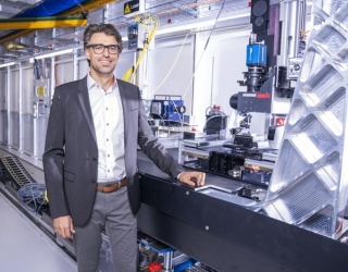
At ECR 2026, Dr. Eliseo Vañó Galván, Head of Radiology at Hospital Nuestra Señora del Rosario in Madrid, discussed why his department implemented Philips’ Verida CT system. In our interview, he explains how spectral CT is improving cardiac imaging, workflow efficiency and radiation dose optimization in daily clinical practice.
You recently implemented Philips’ flagship CT system. What were the conditions in your radiology department before that decision that made you start looking for a new solution?
Dr. Eliseo Vañó Galván: Currently, we have an independent CT and MR department. Until recently, we had two CT scanners. One of them was a Philips Spectral Detector CT 7500, which we have been using since 2021. The other one was an older, conventional 64-slice scanner from another vendor, in this case GE.
We wanted to upgrade that older system. Over the past five years, our experience with spectral detector CT has made us completely dependent on this technology, because we have clearly seen the advantages it brings for our patients. So, we wanted a solution that was at least comparable to the CT 7500, or ideally even better.
On my desk, I had proposals from photon-counting vendors as well as the Philips Verida CT system. It was not an easy decision because all these high-end scanners are excellent systems and the offers were very competitive. In the end, however, I decided to go with Verida CT because I felt it was the most usable system in terms of workflow efficiency, patient throughput, image acquisition, and the management of spectral data.
With photon-counting systems, the spatial resolution is certainly impressive. However, from my perspective, you lose some of the usability and flexibility of spectral functionality. With Verida CT, spectral capabilities are much easier to access and integrate into daily clinical practice.
For example, from the PACS interface you can simply right-click and access spectral data in less than five seconds. That kind of workflow is currently more difficult with photon-counting systems because of the very large data volumes they generate.
We already have five years of experience with spectral CT and have become a reference site, for example for cardiac spectral imaging. In our experience, the advantages for patient outcomes are greater than the additional spatial resolution offered by photon-counting systems. So even though we had strong offers from photon-counting vendors, I ultimately decided that Verida CT was the safest choice.
From a reimbursement and business model perspective, the implementation was also much easier for us. The installation itself was very straightforward. From the moment of installation, it took only about 48 hours before the system was ready for its first scan, and within less than five days the scanner was already being used with patients.
So far, we have already scanned more than 350 patients. We are seeing excellent image quality and radiation dose reductions of up to 70 percent compared to the CT 7500, which was already a very strong spectral CT system. We started from a very good baseline, and now we have achieved even greater radiation dose optimization. Overall, we are very happy with the first results. Our initial impressions are extremely positive, and the image quality is terrific.
Did the new system replace your oldest CT scanner, or do you now operate three units?
Dr. Eliseo Vañó Galván: No, it replaced our conventional 64-slice CT system. Now we operate two systems: the CT 7500 and the new Verida CT. This means we are now a fully spectral CT department. Another important step for us was upgrading the CT 7500 with “Precise Image”, which is Philips’ deep learning–based reconstruction technology. With this upgrade we can achieve excellent image quality while further reducing radiation dose. So today we are not only a fully spectral department, we are also a fully precise-enabled department.
While I am here at the ECR congress, my team in Madrid is already implementing the new protocols on the second scanner, and so far the results look very promising in terms of image quality and dose optimization. I am really looking forward to going back to Madrid to review the results in detail, because I think we are making very significant progress in radiation dose optimization.
You mentioned cardiac imaging. Was that one of the main drivers behind your decision?
Dr. Eliseo Vañó Galván: Yes, it was certainly one of the most important factors. Cardiac imaging is probably the area of radiology that is currently growing the fastest. Cardiovascular disease remains the leading cause of death worldwide, and if you compare the situation to five years ago, cardiac imaging has seen one of the strongest increases in demand. It is also the area with the longest waiting lists for both CT and MR examinations.
This system is particularly well suited for cardiac imaging for several reasons. It can acquire a complete cardiac study in just a couple of heartbeats. That makes the acquisition very straightforward and convenient for both the patient and the radiologist or cardiologist interpreting the study. In addition, it is currently the only energy-integrating detector system that can provide full spectral information, which adds another important diagnostic dimension.