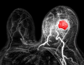
Multiparametric MRI Improves Tumor Differentiation
New research published in Academic Radiology suggests that combining time-dependent diffusion MRI (Td-dMRI) parameters with macromolecular proton fraction (MPF) may improve the differentiation of cervical cancer grades.
The study found that a combination of cellularity, intracellular volume fraction (Vin), and MPF achieved:
- 96% AUC
- 97.14% sensitivity
- 82.46% specificity
These results indicate improved diagnostic performance compared to individual MRI-derived parameters.
Study Design and Patient Cohort
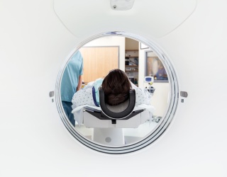
For the prospective study, researchers analyzed imaging data from 92 patients with cervical cancer who underwent Td-dMRI and 3T MPF imaging.
- 57 patients had low-grade (grade I/II) cervical cancer
- 35 patients had high-grade (grade III) cervical cancer
The study evaluated the ability of imaging biomarkers to differentiate tumor grade prior to treatment.
Comparison with Individual MRI Parameters
The study authors reported that individual imaging parameters demonstrated lower diagnostic performance compared to the combined approach:
Cellularity:
- AUC: 92.3%
- Sensitivity: 88.57%
- Specificity: 77.19%
Vin:
- AUC: 85.4%
- Sensitivity: 82.86%
- Specificity: 73.68%
MPF:
- AUC: 84.8%
- Sensitivity: 91.43%
- Specificity: 68.42%
According to the researchers, these findings support the use of a multiparametric MRI approach for improved tumor grading.
Imaging Biomarkers Reflect Tumor Biology
The study authors noted that Td-dMRI-derived parameters such as cellularity and Vin are associated with tumor characteristics and may support non-invasive grading.
“An important explanation for this outcome may be the greater aggressiveness associated with high-grade lesions. Specifically, highly aggressive lesions exhibit enhanced cell proliferation—a process that directly induces an increase in cell density and intracellular volume fraction, while simultaneously reducing extracellular space. Further, incomplete cellular maturation and intercellular compression, driven by this rapid proliferation, also lead to a decrease in cell diameter. Collectively, these pathological changes ultimately result in elevated cellularity and Vin, as well as reduced diameter and Dex, (extracellular diffusivity),” noted lead study author Nan Meng, MD, , who is affiliated with the Department of Radiology at Henan Provincial People’s Hospital and Zhengzhou University People’s Hospital in Zhengzhou, China, and colleagues.
Role of MPF in Cervical Cancer Assessment
The researchers also found that MPF provided independent value in differentiating tumor grades.
“Results showed that MPF values in the high-grade group were significantly higher than those in the low-grade group … We hypothesize that differences in collagen deposition across CC with varying malignancy grades may serve as a primary factor contributing to these observed MPF alterations. Additionally, tumor stromal proliferation and cellular death may also play a role in driving this change,” added Meng and colleagues.
Study Limitations
The authors acknowledged several limitations, including:
- Single-center study design
- Relatively small patient cohort
- Lack of external validation
- Exclusion of lesions smaller than 1 cm due to spatial resolution constraints
Implications for Preoperative MRI Assessment
The findings suggest that combining Td-dMRI parameters with MPF may support improved preoperative differentiation between low-grade and high-grade cervical cancer.
Source: Academic Radiology