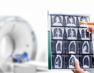
What was once used mainly to evaluate chest pain is now emerging as a foundation for early disease detection, personalized prevention, and, in the future, even screening people before symptoms ever appear.
A major catalyst for this shift came in 2021, when a coalition of leading medical societies elevated CCTA to a Class I, first-line recommendation for patients with stable, intermediate-risk chest pain. This wasn’t just another guideline update—it was a signal that the field had irreversibly changed. CCTA became recognized among the most accurate non-invasive tools for diagnosing coronary disease, and adoption accelerated across the country. CCTA volumes rose sharply, health systems began redesigning chest pain pathways around it, and demand grew rapidly for non-invasive, image-based decision-support technologies.
The 2021 Chest Pain Guideline effectively transformed CCTA from an optional test into the new first-line standard for diagnosing coronary disease—driving adoption, reimbursement, technology innovation, and laying the groundwork for preventive and ultimately asymptomatic cardiovascular care. (Gulati M, Levy PD, Mukherjee D, et al. 2021 AHA/ACC/ASE/CHEST/SAEM/SCCT/SCMR Guideline for the Evaluation and Diagnosis of Chest Pain. Circulation. 2021;144(22):e368–e454)
This momentum also opened the door for reimbursement expansion. Between 2021 and 2024, CCTA became embedded in first-line chest pain pathways, with Medicare and commercial reimbursement increasing and stabilizing in 2024. An adjunct AI technology, FFR-CT, secured Category I CPT status—a permanent U.S. reimbursement designation indicating a clinically established procedure eligible for routine payment. An additional adjunct biomarker, quantitative coronary plaque analysis (QCPA), gained traction and achieved permanent Category I CPT reimbursement, with nationwide coverage beginning in January 2026.
These shifts are transforming cardiac CT from an expensive niche exam into a sustainable, scalable service line—a tool hospitals can now use to build comprehensive chest pain and prevention programs for both symptomatic and, eventually, asymptomatic patients.
So today, the question is no longer if CCTA will become the backbone of preventive cardiac care. The question is when we will fully embrace its power—to detect disease early, guide treatment with precision, and identify high-risk individuals long before the first warning sign.
The Scale of the Problem: Why We Must Evolve
Cardiovascular disease remains the leading cause of death worldwide, with Coronary Artery Disease (CAD) its most fatal form. In the United States, roughly 805,000 people experience a heart attack each year, according to the Centers for Disease Control (CDC), and many are diagnosed far too late—often after plaque has ruptured or progressed to a critical obstruction.
Despite tremendous advances in therapeutics and intervention, the majority of healthcare systems still lack scalable mechanisms to detect CAD early, quantify its biology, monitor its trajectory, and tailor preventive therapy. CCTA—especially when paired with emerging AI biomarkers—may finally offer a pathway to altering disease trajectory at a population level.
From Stress Tests to High-Resolution Coronary Anatomy
For decades, CAD evaluation relied on stress ECG, stress echocardiography, and nuclear perfusion imaging. While clinically useful, these modalities vary in sensitivity and specificity, lack consistency, and provide no direct visualization of coronary anatomy or plaque biology.
CCTA, by contrast, provides high-resolution anatomic detail—revealing not only the presence of stenosis but how plaque disease is distributed and structured along each vessel.
Yet CCTA needs support from additional decision-support tools to determine whether a lesion restricts blood flow or whether plaque is biologically active or rupture-prone. These gaps catalyzed the next wave of AI-enabled innovation.
AI Biomarkers: FFR-CT, Plaque, and Inflammation
Functional Flow: The Rise of FFR-CT – Disease of the Lumen
FFR-CT—pioneered by HeartFlow—was the first major step beyond anatomy.
Built on more than a decade of solid clinical evidence, it estimates fractional flow reserve non-invasively from standard CCTA images, clarifying whether a stenosis meaningfully restricts coronary blood flow.
As demonstrated at RSNA 2025, several companies now offer deep-learning CT-derived physiological or ischemia-assessment solutions —including HeartFlow, Cleerly, Keya Medical, Shukun Technology (now rebranded as Careverse), and Spimed-AI. Some platforms are FDA-cleared; others are approaching submission with strong clinical evidence. By non-invasively quantifying blood flow, these AI tools refine cath-lab triage and more accurately assess qualification for interventions, thereby avoiding unnecessary invasive angiography. Still, physiology alone offers limited insight into the 360° view of a patient’s coronary artery disease. It does not determine disease extent or whether plaque is stable or vulnerable.
Beyond Stenosis: Plaque and Inflammation (Disease of the Vessel Wall and the “Invisible” Disease)
AI-enabled plaque characterization was one of the dominant themes at RSNA 2025.
Several pivotal trials have reshaped our understanding of why plaque—not just stenosis—matters in coronary artery disease. In the emerging field of plaque quantification—referred to as AI-Quantitative Coronary Tomography (AI-QCT) in technical and reimbursement documentation and more recently described by the American College of Cardiology (ACC) as quantitative coronary plaque analysis (QCPA)—the SCOT-HEART trial stands out as a defining milestone.
SCOT-HEART was a pivotal inflection point in establishing the importance of non-obstructive disease. The trial showed that CCTA detects coronary artery disease—including non-obstructive plaque frequently missed by stress testing—and that acting on this information with preventive therapy may lead to a durable reduction in myocardial infarction over five years. By demonstrating that visualizing underlying disease biology, not just ischemia, changes outcomes, SCOT-HEART laid the clinical foundation for today’s focus on plaque assessment. SCOT-HEART Investigators. 5-year outcomes of coronary CT angiography in patients with suspected angina due to coronary heart disease. N Engl J Med. 2018;379(10):924–933. doi:10.1056/NEJMoa1805971.
Since SCOT-HEART, the science of coronary plaque has advanced substantially. We now understand that calcified plaque is often relatively stable, whereas lipid-rich, inflamed plaque is far more prone to rupture—highlighting the need for precision-based strategies that target biologically vulnerable disease rather than stenosis alone.
Driven by expanding clinical evidence, favorable reimbursement, and a growing emphasis on prevention, a wave of plaque-focused companies and technologies has emerged, including Cleerly, HeartFlow, Circle Cardiovascular, syngo® CT Coronary Cockpit by Siemens Healthineers, Medis, Caristo, Keya Medical, and Shukun—some FDA-cleared, others approaching regulatory submission.
Modern plaque tools quantify total burden, differentiate plaque subtypes, and identify high-risk morphologic features—shifting evaluation beyond luminal narrowing and toward understanding the underlying biology of disease. Contemporary AI-QCT platforms have the potential to enable clinicians to detect vulnerable plaque, monitor therapeutic response, and personalize care based on biological risk rather than stenosis alone.
This marks a fundamental shift in cardiovascular medicine: from AI guiding decisions primarily for intervention to AI guiding decisions for treatment and prevention.
Inflammation: The Added Dimension
Coronary inflammation imaging—especially perivascular fat attenuation metrics—adds a new dimension to cardiovascular risk assessment. A landmark Lancet study demonstrated that perivascular fat attenuation (fat attenuation index, FAI) measured on routine CCTA strongly predicts future cardiac events even in patients without obstructive CAD (Oikonomou EK et al., The Lancet 2018;392:929–939, https://doi.org/10.1016/S0140-6736(18)31114-0).
Notably, perivascular FAI (fat attenuation index) scoring identified a subset of patients who appeared disease-free by traditional measures but had markedly higher inflammatory risk, associated with a substantially higher long-term risk of cardiac mortality.
Although not yet FDA cleared and not ready for routine clinical use, this biomarker holds substantial promise for detecting “invisible” disease even before plaque formation.
Together, plaque and emerging inflammation analytics provide a more complete risk profile and position CCTA as a powerful tool for therapeutic targeting, longitudinal monitoring, and early preventive intervention.

Technology Tailwinds: Photon-Counting CT (PCCT) Hardware Innovation
A critical determinant of accurate CAD quantification lies in the underlying scanner technology. The arrival of photon-counting CT (PCCT), showcased prominently across RSNA 2025, represents one of the most transformative hardware advances in cardiac imaging in decades. Unlike conventional energy-integrating detectors, PCCT captures individual photons with greater precision, enabling markedly enhanced spatial resolution, improved contrast-to-noise ratios, and sharper visualization of subtle plaque characteristics. These capabilities directly impact the fidelity of plaque and inflammation assessment—domains where minor imaging variations can meaningfully influence clinical interpretation.
Recent evidence underscores this impact. A large retrospective study of nearly 8,000 patients demonstrated that dual-source photon-counting CT (PCCT) substantially improved diagnostic performance compared with traditional energy-integrating detector CT systems, resulting in fewer unnecessary referrals for invasive coronary angiography. Importantly, when patients were referred based on PCCT findings, they were more likely to undergo revascularization—suggesting superior identification of clinically actionable disease (Sakai K, Shin D, Singh M, et al. J Am Coll Cardiol. 2025;85(4):339–348. doi:10.1016/j.jacc.2024.10.069).
As Bernhard Schmidt, PhD Head of CT Innovation, Physics and Global Collaboration at Siemens Healthineers, emphasized during RSNA 2025:
“PCCT is a paradigm shift. We are beginning to see early diseases that would appear normal under EID-CT (i.e. traditional energy-integrating detector CT). This has profound implications for early diagnosis, therapy response, and reproducibility. In the next 5–10 years, the field will transition from energy-integrating detectors to photon-counting technology.”
Siemens Healthineers’ launch of the NAEOTOM Alpha in 2021—the first commercially available dual-source photon-counting CT (PCCT) system—marked a major milestone in cardiac imaging. Other major vendors are expected to enter the market with PCCT solutions in the near future. In parallel, CT manufacturers and AI vendors are already validating their algorithms on PCCT datasets to ensure consistent and reproducible AI biomarker quantification as clinical adoption accelerates.
Designing With the End Game in Mind
Remaining Adoption Challenges
Despite strong technological, clinical, and economic tailwinds—and a projected CCTA CAGR above 25% over the next five years—health systems still face substantial adoption barriers. Many centers struggle with profitability, staffing, and the legacy dominance of reimbursed nuclear imaging. Institutions require education and operational support to implement CCTA workflows successfully—an effort championed by SCCT, but still uneven across sites.
Plaque analysis tools exhibit substantial variability in both output quality and reproducibility, as detailed in the 2024 expert consensus statement from the Society of Cardiovascular Computed Tomography (SCCT) addressing quantitative CCTA standards (https://pubmed.ncbi.nlm.nih.gov/38849237).
Bernhard Schmidt emphasized the need for an industry-wide standardization effort: consistent reconstruction kernels, harmonized metrics, and validated scoring frameworks—similar to early calcium-scoring standardization. Without reproducibility across vendors, advanced plaque and inflammation biomarkers cannot reliably be adopted into therapeutic disease management.
Schmidt’s comments were echoed in recent American College of Cardiology (ACC) consensus guidance on quantitative coronary plaque analysis using CCTA, which acknowledged the technology’s potential to refine cardiovascular risk assessment while emphasizing that its role in clinical decision-making remains adjunctive and constrained by current evidence gaps. (JACC: Cardiovascular Imaging, ACC Expert Consensus Statement, January 2026. Consensus statement: https://www.jacc.org/doi/10.1016/j.jcmg.2025.11.008)
A more detailed analysis of the ACC panel’s findings—including implications for serial imaging, workflow standardization, and clinical adoption—will be presented in a forthcoming article.
Core-Lab Dependence and the Scalability Imperative
Most AI platforms we saw at RSNA 2025 still rely on human-in-the-loop core-lab models with turnaround times measured in hours. That paradigm cannot support the growing case volumes anticipated in the coming years—certainly not the urgent demands of emergency department triage, where rapid decision-making is essential for reducing the high costs associated with heart attack evaluation. And it is even less compatible with the scale required for population-level cardiovascular screening of tens of millions of prospective patients.
At the same time, health systems increasingly want control over their data, workflows, and turnaround expectations. This is pushing the field toward on-premise, autonomous AI solutions, placing analytical control directly in the hands of the site. To meet these demands, AI must evolve into solutions that are automated, reproducible, and natively integrated into scanners, PACS, and EMR workflows.
Today’s commercial platforms still require meaningful human assistance; algorithms must improve to minimize this dependency. Only then can we deliver a truly scalable infrastructure capable of supporting the future of cardiovascular screening.
As Dr. Paul, CEO of Spimed-AI French start-up, noted at RSNA 2025: “Autonomous FFR-CT and plaque platforms will be essential for scaling CCTA from chest pain evaluation to population-level screening. This brings the technology into the hands of the institutions that own the patient data and can fully control their workflow around deploying AI.”
Success Recipe & Future Outlook
From Blockages to Biology, From Crisis to Early Detection & Prevention
RSNA 2025 signaled that the coming year may be a true tipping point, with simultaneous momentum around FFR-CT, AI-QCT, and emerging inflammation tools. While CCTA today is primarily used for symptomatic chest pain and cath lab triage, its greatest long-term opportunity lies in the early detection of cardiovascular disease—and eventually in high-risk screening of asymptomatic patients, much like mammography or colonoscopy. Recent ACC consensus guidance reinforces both the promise of this shift and the need for careful, evidence-driven adoption as these technologies mature.
Eric J. Topol, MD, a leading cardiologist–scientist and widely recognized thought leader in cardiovascular medicine, patient empowerment and digital health, and the bestselling author of The Creative Destruction of Medicine, The Patient Will See You Now, and Deep Medicine, has described a “big shift” in cardiology toward the detection of early atheroma and arterial inflammation—moving beyond luminal stenosis to interrogate the underlying biology driving disease progression. His central question remains at the heart of the field: How can these advanced imaging approaches be made accessible, affordable, and scalable to support population-level prevention? (Topol EJ. The Big Shift in Cardiology to Atheroma and Inflammation. Ground Truths. November 16, 2025. https://erictopol.substack.com/p/the-big-shift-in-cardiology-to-atheroma)
Meeting that challenge requires designing technologies for the future rather than the present. AI systems must improve autonomous performance and reduce reliance on human work-up. Vendors must collaborate on reproducible, cross-vendor standards so that plaque and inflammation measurements are harmonized across platforms. Companies need to think beyond today’s symptomatic pathways and build infrastructures capable of supporting tens of millions of annual scans, with deep integration across scanners, PACS, and patient coordination systems. Professional societies will also need to guide the development of screening frameworks once sufficient prospective evidence is established.
Without designing with the end game in mind, even the most sophisticated tools will ultimately reach only a fraction of the patients who could benefit and will never meaningfully alter patient management.

Cardiac CTA has evolved from a diagnostic exam into a cornerstone of comprehensive coronary assessment. We now stand at the threshold of a transformation that will reshape cardiovascular care—from fixing blockages to understanding plaque biology; from episodic imaging to longitudinal tracking; and from reactive treatment to proactive, population-level prevention.
The enabling technologies are here. Reimbursement is improving. Guidelines are evolving. Momentum is accelerating - and now with clearer guardrails.
When we succeed, the future of cardiac care will look fundamentally different: asymptomatic individuals undergoing standardized, non-invasive CCTA screening; plaque and inflammation monitored longitudinally; and preventive therapies personalized long before a first event occurs. The opportunity—in clinical outcomes, economic value, and human lives—is immense. And as RSNA 2025 clearly demonstrated, the work to build this future is already underway.
Industry Expert Series written by Andjela Azabagic