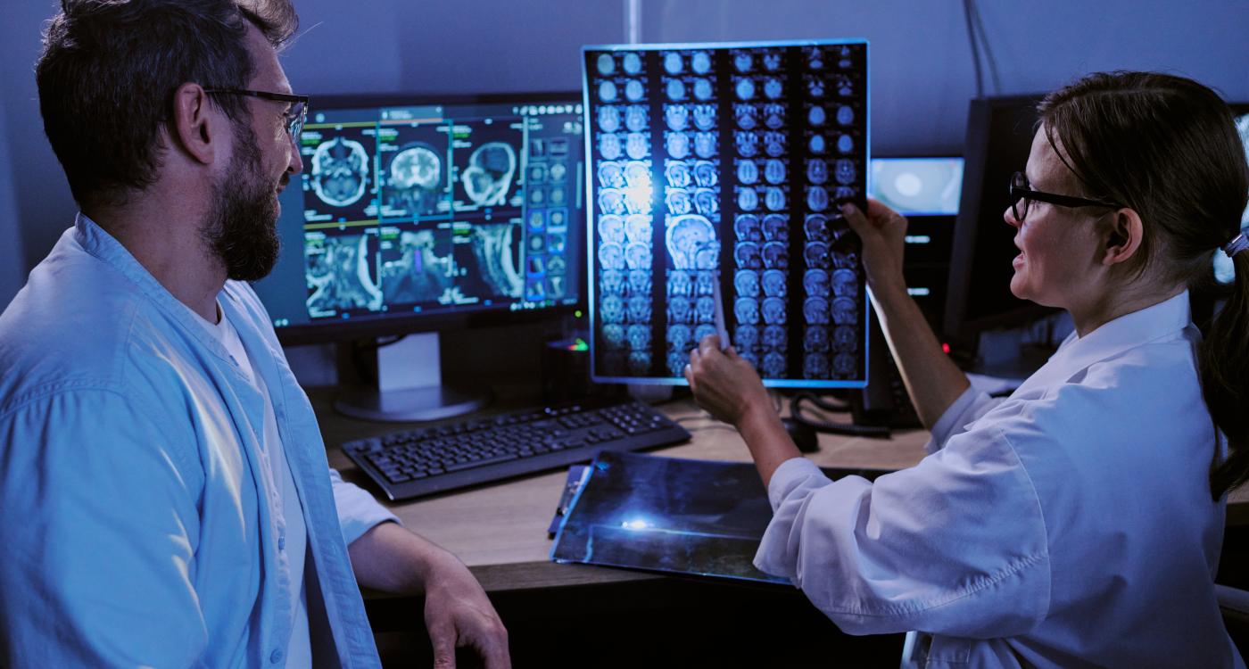
Digital twins in medicine are no longer conceptual visions of future healthcare. In cardiovascular imaging, oncology, and pulmonary medicine, patient-specific computational models are already influencing real clinical decisions.
At RSNA 2025, experts discuessed how digital twins are evolving from research tools into decision-support systems embedded in practice. The session “Digital Twins in Medicine: Clinical Applications and the Fundamental Role of Imaging” brought together computational scientists and clinical leaders, moderated by Kenneth C-Y. Wang, MD, PhD and Summer J. Decker, PhD, and featuring Charles Taylor, PhD, co-founder of HeartFlow and Moncrief Chair in Computational Medicine at UT Austin, and Caroline Chung, MD, Vice President and Chief Data and Analytics Officer and co-director of the Institute for Data Science in Oncology at MD Anderson Cancer Center.
The central concept of digital twins is straightforward yet ambitious: create a virtual model of a biological system that is continuously updated with patient data and capable of predicting outcomes.
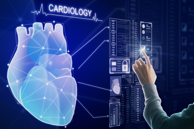
Digital Twins in Cardiology
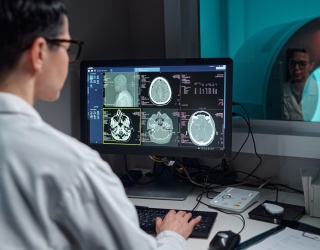
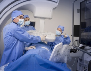
The most established clinical example is coronary CT–derived fractional flow reserve (FFRCT), integrating anatomy, physiology, and increasingly, plaque characterization. The clinical question driving its development was straightforward: could obstructive coronary disease be identified before a patient enters the cardiac catheterization laboratory?
“If you're trying to diagnose coronary artery disease,” Taylor noted, “you really do need to look at the coronary arteries”.
By extracting coronary anatomy from CT angiography, applying physiologic boundary conditions, and solving fluid dynamics equations, digital models compute pressure gradients along the coronary tree. This transforms CT from a purely anatomical modality into a functional assessment tool.
While AI supports segmentation and data processing, Taylor emphasized the underlying foundation: “Quite frankly, there’s no substitute for a little bit of physics,” he said.
Today, CT-based physiology modeling allows clinicians to:
- Defer invasive catheterization when physiology is normal
- Identify lesion-specific ischemia
- Guide revascularization decisions
Digital twin applications in cardiology now extend beyond flow simulation. Quantitative coronary plaque analysis enables separation of calcified and non-calcified plaque and identification of high-risk phenotypes. When plaque characterization is combined with physiology modeling, risk prediction improves significantly compared to percent stenosis alone. This integration of anatomy, plaque phenotype, and hemodynamics reflects the broader promise of digital twins: merging structural and functional data into a predictive framework. “It’s far better for an interventional cardiologist to walk into a procedure knowing as much as possible about what they’re going to do,” Taylor said.

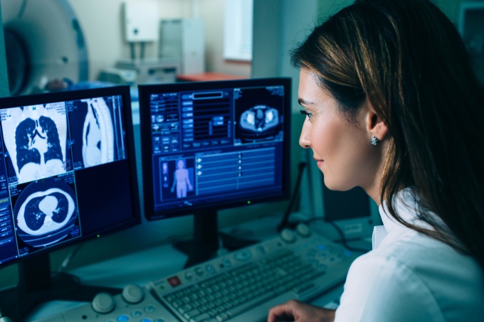
Lung Modeling and Simulation-Based Ventilation
Digital twin concepts are also expanding into pulmonary medicine. CT-derived airway models allow simulation of mechanical ventilation parameters and inhaled therapeutics to identify better drug delivery for patients with idiopathic pulmonary fibrosis. By exploring parameter adjustments virtually, clinicians may also optimize oxygenation strategies and reduce the risk of ventilator-induced lung injury. Thus, imaging provides the structural map, while computational modeling delivers predictive insights.
Oncology: Adaptive Therapy and Continuous Model Updating
In oncology, digital twins introduce greater complexity — and opportunity.
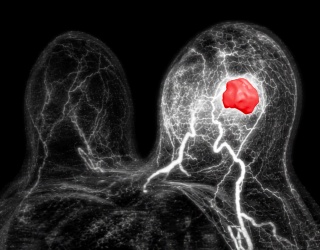
Caroline Chung emphasized the need for models to be “fit for purpose.” In cancer care, digital twins incorporate multiparametric MRI, diffusion-weighted imaging, and treatment data to simulate patient and tumor response over time. Unlike static imaging snapshots, these models evolve during therapy. Imaging, blood markers, and treatment data continuously feed into the virtual model, which in turn simulates potential outcomes and informs therapy decisions.
Using glioblastoma as an example, Chung described how repeated imaging could recalibrate tumor growth models and potentially predict progression before symptoms occur. “There are multiple clinical trials open that are looking at leveraging diffusion-weighted imaging to actually inform whether the patient's actually responding, adapting treatment, potentially up to even on a daily basis.” This creates the possibility of adaptive radiotherapy, adjusting treatment before visible progression occurs.
However, oncology decisions are complex and rarely binary. Chung noted, that human decision-making under uncertainty, involves both physician and patient, and must account for quality-of-life considerations. That makes transparency critical. "It's not to say that we can get to an uncertainty of zero. That is not the intention. It's about being informed about what that uncertainty is," she said.
Imaging as the Structural Backbone
Across specialties, one theme was consistent: digital twins depend on reliable imaging data. Imaging is not merely an input variable; it is the structural backbone of predictive medicine.
The session highlighted that digital twins are already used in cardiovascular care and are advancing in oncology medicine. Successful implementation requires:
- High-quality, standardized imaging
- Physics-informed modeling
- AI-assisted data integration
- Clinical validation
- Integration into clinical workflows
As discussed through our digital twin series and at RSNA 2025, digital twins represent an evolution of quantitative imaging rather than a replacement of clinical expertise. By linking imaging, computational modeling, and predictive analytics, they aim to support more informed, patient-specific decisions across multiple specialties.
Digital Twins Series